Cervical Spondylosis
The neck is part of a long flexible column, known as the spinal column or backbone, which extends through most of the body. The cervical spine (neck region) consists of seven bones (C1-C7 vertebrae), which are separated from one another by intervertebral discs. These discs allow the spine to move freely and act as shock absorbers during activity. (1)
What does the cervical spine do?
Your cervical spine has several functions, including: (2)
- Protecting your spinal cord. The nerves of your spinal cord pass through a large hole (called the vertebral foramen) that passes through the center of all of your vertebrae — from the base of your skull through the cervical vertebrae, the thoracic (middle back) vertebrae and ending between the first and second lumbar (lower back) vertebrae. Taken together, all the stacked vertebrae of your spine form a protective central canal that protects your spinal cord.
- Supporting your head and allowing movement. Your cervical spine supports the weight of your head (average weight of 10 to 13 pounds). It also allows your head and neck to tilt forward (flexion), backward (extension), turn from side to side (rotation), or bend to one side (ear-to-shoulder; lateral flexion).
- Providing a safe passageway for vertebral arteries. Small holes in cervical spine vertebrae C1 to C6 provide a protective pathway for vertebral arteries to carry blood to your brain. This is the only section of vertebrae in the entire spine that contains holes in the bone to allow arteries to pass through.
Cervical spondylosis
Cervical spondylosis (Arthritis of the Neck) is a general term for age-related wear and tear affecting the spinal disks in your neck. As the disks dehydrate and shrink, signs of osteoarthritis develop, including bony projections along the edges of bones (bone spurs). (3)
Cervical spondylosis is very common and worsens with age. More than 85% of people older than age 60 are affected by cervical spondylosis.
Symptoms
Most people experience no symptoms. When symptoms do occur, they typically include pain and stiffness in the neck.
Sometimes, cervical spondylosis results in a narrowing of the spinal canal within the bones of the spine (the vertebrae). The spinal canal is the space inside the vertebrae that the spinal cord and the nerve roots pass through to reach the rest of the body. If the spinal cord or nerve roots become pinched, you might experience: :(3)
- Pain in the neck that may travel to your arms or shoulders
- Headaches
- A grinding feeling when you move your neck
- Weakness in your arms and legs
- Numbness in your shoulders, arms, or hands
- Stiff neck
- Trouble keeping your balance, especially when walking
- Trouble controlling your bladder or bowels

Causes of cervical spondylosis
As you get older, your spine changes due to decades of normal wear and tear. Starting in middle age, the disks between your vertebrae start to change. These changes can include: (4)
- Degeneration: The spinal disks in your neck may slowly wear down (degenerate). With time, the disks become thinner, and the soft tissue has less elasticity. If you or your parents measure in a little shorter in height than you did years ago, this is normal collapsing or settling of your disks.
- Herniation: Normal aging can cause part of your spinal disk to tear or crack. This is called a herniated disk. The herniation can allow the disk to bulge out, pressing on nearby tissue or a spinal nerve. This pressure can cause pain, tingling, or numbness.
- Osteoarthritis: Osteoarthritis is a progressive (ongoing) condition that causes the cartilage in your joints to degenerate (wear down with time). With osteoarthritis, cartilage degenerates faster than normal aging.
- Bone spurs: When cartilage in the joints of the vertebrae in your spine starts to degenerate and bone tissue rubs directly against other bone tissue, abnormal bone growths develop along the edges of vertebrae. These growths (called osteophytes or bone spurs) are common as you age. Often, they cause no symptoms.
Diagnosis
Your healthcare provider will likely start with a physical exam that includes:(5) (6)
- Checking the range of motion in your neck
- Testing your reflexes and muscle strength to find out if there's pressure on your spinal nerves or spinal cord
- Watching you walk to see if spinal compression is affecting your gait
Imaging tests
Imaging tests can provide detailed information to guide diagnosis and treatment. Examples include:
- Neck X-ray. An X-ray can show changes in the spine, such as bone spurs, that indicate cervical spondylosis. Neck X-rays can also rule out rare and more serious causes of neck pain and stiffness, such as tumors, cancer, infections, or fractures.
- MRI. Using radio waves and a strong magnetic field, MRI can produce detailed images that can help pinpoint areas where nerves might be pinched.
- CT myelography. In this type of CT scan, a dye is injected into the spinal canal to provide more-detailed imaging. This test makes it easier to see the details of the spinal cord, spinal canal, and nerve roots.
Nerve function tests
You may need tests to determine if nerve signals are traveling properly to your muscles. Nerve function tests include:
- Electromyography. This test measures the electrical activity in your nerves as they transmit messages to your muscles when the muscles are contracting and at rest.
- Nerve conduction study. Electrodes are attached to the skin above the nerve to be studied. A small shock is passed through the nerve to measure the strength and speed of nerve signals
Treating cervical spondylosis
Treatments for cervical spondylosis focus on providing pain relief, lowering the risk of permanent damage, and helping you lead a normal life.
Nonsurgical methods are usually very effective.
Physical therapy
Your doctor might send you to a physical therapist for treatment. Physical therapy helps you stretch your neck and shoulder muscles. This makes them stronger and ultimately helps to relieve pain. (7)
You might also have neck traction. This involves using weights to increase the space between the cervical joints and relieve the pressure on the cervical discs and nerve roots.
Medications
Your doctor might prescribe certain medications if over-the-counter (OTC) drugs don’t work. These include: (7)
- muscle relaxants, such as cyclobenzaprine, to treat muscle spasms
- narcotics, such as hydrocodone, for pain relief
- anti-epileptic drugs, such as gabapentin, to relieve pain caused by nerve damage
- steroid injections, such as prednisone, to reduce tissue inflammation and subsequently lessen pain
- prescription nonsteroidal anti-inflammatory drugs (NSAIDs), such as diclofenac (Voltaren-XR), to reduce inflammation
Surgery
If your condition is severe and doesn’t respond to other forms of treatment, you might need surgery. This can involve removing bone spurs, parts of your neck bones or herniated discs to give your spinal cord and nerves more room.
Surgery is rarely necessary for cervical spondylosis. However, a doctor may recommend it if the pain is severe and it’s affecting your ability to move your arms.
Home treatment options
If your condition is mild, you can try a few things at home to treat it: (7)
- Take an OTC pain reliever, such as acetaminophen or an NSAID, which includes ibuprofen and naproxen sodium.
- Use a heating pad or a cold pack on your neck to provide pain relief for sore muscles.
- Exercise regularly to help you recover faster.
- Wear a soft neck brace or soft collar to get temporary relief. However, you shouldn’t wear a neck brace or collar for long periods because that can make your muscles weaker.
Makkah Pharmacy recommends:


5 easy-to-follow at-home exercises to reduce cervical spondylosis pain: (8)
Push forward
This exercise is very easy to follow and also helps better flexibility in the neck. Here's how to perform the neck push-forward exercise:
- Stand straight
- Try to push your neck forward as far as possible
- At this point, your neck would be protruding forward ahead of the rest of your body
- Hold this stretch for a few snoods and release
- Repeat it a few times daily
Neck tilts
Neck tilts as the name suggests helps in stretching the neck in different directions and help relieve pain and stiffness. Here is how you can practice neck tilts.
- Try to place your head on your right shoulder
- At this point, your right ear should be touching your right shoulder
- Hold this position for a few seconds
- Now witch, with your head resting on your left shoulder
- Repeat this at least 4-5 times, a few times a week, or whenever you feel discomfort
Neck bowing
Similar to the pushing the neck forward exercise, neck bowing focuses on stretching the neck out as far as possible. A combination of these two regularly can ensure better flexibility and a reduction in pain in the neck. Here's how you perform the neck-bowing exercise:
- Try to rest your neck on your chest
- In this position, your chin should be touching your chest
- Hold this position for a few seconds and release
- Repeat this along with push forward exercise, alternatively
- Repeat at least 3-4 times
Look behind
As the name suggests, the looking behind neck exercise focuses on stretching the neck behind. For this exercise, the motive is to turn your neck around in an attempt to see behind you. Here's how to practice the look behind the exercise:
- Stand straight and turn your neck to the left as far behind as you comfortably can
- Hold for a few seconds
- Repeat on the other side
- Repeat this set 4-5 times or more, however comfortable
- Please note that your body must stay in the same position and angle throughout
Raised shoulders
This is another easy-to-follow exercise for cervical spondylosis. It also helps release tension from the neck and promotes a calming feeling. This is how to practice raised shoulders:
- As the name suggests, try to raise your shoulders as you normally would
- At this point, your shoulders might be touching your ears
- Hold this position for a few songs and release
- You can practice this position 10-15 times daily to reduce pain and stiffness
References:
1. Cervical Spine – Anatomy, Diseases, and Treatments [Internet]. [cited 2023 Feb 21]. Available from: https://www.aans.org/en/Patients/Neurosurgical-Conditions-and-Treatments/Cervical-Spine
2. Cervical Spine (Neck): What It Is, Anatomy & Disorders [Internet]. [cited 2023 Feb 21]. Available from: https://my.clevelandclinic.org/health/articles/22278-cervical-spine
3. Cervical spondylosis - Symptoms and causes - Mayo Clinic [Internet]. [cited 2023 Feb 21]. Available from: https://www.mayoclinic.org/diseases-conditions/cervical-spondylosis/symptoms-causes/syc-20370787
4. Cervical Spondylosis: Symptoms, Causes, Treatments [Internet]. [cited 2023 Feb 21]. Available from: https://my.clevelandclinic.org/health/diseases/17685-cervical-spondylosis
5. Theodore N. Degenerative Cervical Spondylosis. New England Journal of Medicine. 2020 Jul 9;383(2):159–68.
6. Cervical spondylosis - Diagnosis and treatment - Mayo Clinic [Internet]. [cited 2023 Feb 21]. Available from: https://www.mayoclinic.org/diseases-conditions/cervical-spondylosis/diagnosis-treatment/drc-20370792
7. Cervical Spondylosis: Causes, Symptoms, Home Treatments, and More [Internet]. [cited 2023 Feb 21]. Available from: https://www.healthline.com/health/cervical-spondylosis#treatment
8. Cervical Spondylosis: 5 Exercises To Reduce Neck Pain [Internet]. [cited 2023 Feb 21]. Available from: https://www.ndtv.com/health/cervical-spondylosis-5-exercises-to-reduce-neck-pain-2988112
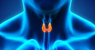
Thyroid
The thyroid gland is a small organ that’s located in the front of the neck, wrapped around the windpipe (trachea). It’s shaped like a butterfly, smaller in the middle with two wide wings that extend around the side of your throat. The thyroid is a gland. You have glands throughout your body, where they create and release substances that help your body do a specific thing. Your thyroid makes hormones that help control many vital functions of your body. (1)
What does the thyroid do?
Your thyroid has an important job to do within your body — releasing and controlling thyroid hormones that control metabolism. Metabolism is a process where the food you take into your body is transformed into energy. This energy is used throughout your entire body to keep many of your body’s systems working correctly. Think of your metabolism as a generator. It takes in raw energy and uses it to power something bigger. (1)
The thyroid controls your metabolism with a few specific hormones — T4 (thyroxine, which contains four iodine atoms) and T3 (triiodothyronine, which contains three iodide atoms). These two hormones are created by the thyroid and they tell the body’s cells how much energy to use. When your thyroid works properly, it will maintain the right amount of hormones to keep your metabolism working at the right rate. As the hormones are used, the thyroid creates replacements. (1)
This is all supervised by something called the pituitary gland. Located in the center of the skull, below your brain, the pituitary gland monitors and controls the amount of thyroid hormones in your bloodstream. When the pituitary gland senses a lack of thyroid hormones or a high level of hormones in your body, it will adjust the amounts with its hormone. This hormone is called thyroid-stimulating hormone (TSH). The TSH will be sent to the thyroid and it will tell the thyroid what needs to be done to get the body back to normal.
Thyroid disease is a general term for a medical condition that keeps your thyroid from making the right amount of hormones. Your thyroid typically makes hormones that keep your body functioning normally. When the thyroid makes too much thyroid hormone, your body uses energy too quickly. This is called hyperthyroidism. Using energy too quickly will do more than make you tired — it can make your heart beat faster, cause you to lose weight without trying, and even make you feel nervous. On the flip side of this, your thyroid can make too little thyroid hormone. This is called hypothyroidism. When you have too little thyroid hormone in your body, it can make you feel tired, you might gain weight and you may even be unable to tolerate cold temperatures.

Hyperthyroidism (overactive thyroid)
Symptoms
Hyperthyroidism sometimes looks like other health problems. That can make it hard to diagnose. It can cause many symptoms, including:(2,3)
- Losing weight without trying.
- Fast heartbeat, a condition called tachycardia.
- Irregular heartbeat, also called arrhythmia.
- Pounding of the heart, sometimes called heart palpitations.
- Increased hunger.
- Nervousness, anxiety, and irritability.
- Tremor, usually a small trembling in the hands and fingers.
- Sweating.
- Changes in menstrual cycles.
- Increased sensitivity to heat.
- Changes in bowel patterns, especially more-frequent bowel movements.
- Enlarged thyroid gland, sometimes called a goiter, which may appear as a swelling at the base of the neck.
- Tiredness.
- Muscle weakness.
- Sleep problems.
- Warm, moist skin.
- Thinning skin.
- Fine, brittle hair.
Causes
Many diseases and conditions can cause hyperthyroidism, including:(4)
- Graves’ disease (the most common cause of hyperthyroidism)
- Inflammation (thyroiditis) of the thyroid due to viral infections, some medicines, or after pregnancy (common)
- Taking too much thyroid hormone (common)
- Noncancerous growths of the thyroid gland or pituitary gland (rare)
- Some tumors of the testes or ovaries (rare)
- Getting medical imaging tests with contrast dye that has iodine (rare, and only if there is a problem with the thyroid)
- Eating too many foods that contain iodine (very rare, and only if there is a problem with the thyroid)
Exams and Tests
The healthcare provider will do a physical exam. The exam may find the following:(5)
- High systolic blood pressure (the first number in a blood pressure reading)
- Increased heart rate
- Enlarged thyroid gland
- Shaking of the hands
- Swelling or inflammation around the eyes
- Very strong reflexes
- Skin, hair, and nail changes
Blood tests are also ordered to measure your thyroid hormones TSH, T3, and T4.
You may also have blood tests to check:
- Cholesterol levels
- Glucose
- Specialized thyroid tests like Thyroid receptor antibody (TRAb) or Thyroid Stimulating Immunoglobulin (TSI)
Imaging tests of the thyroid may also be needed, including:
- Radioactive iodine uptake and scan
- Thyroid ultrasound (rarely)
Risk factors
The main risk factors for hyperthyroidism are:(6)
- Sex. Females are much more likely to have hyperthyroidism than males. Experts believe this may have to do with hormones.
- Pregnancy. Pregnancy can stimulate hyperthyroidism in some people, which can cause complications for both the parent and fetus.
- Age. You’re considered at higher risk for hyperthyroidism as an older adult, especially after age 60.
- Genetics. A family history of hyperthyroidism usually indicates an increased likelihood of developing the condition.
- Iodine exposure. You may get too much iodine from certain medications or foods.
- Having another health condition. People with type 1 diabetes, primary adrenal insufficiency, or pernicious anemia are considered more at risk.
Lifestyle is an important part of lowering your risk factors for hyperthyroidism. This includes eating a balanced diet, taking nutritional supplements if necessary, and getting adequate physical activity during the week. Work with your doctor to create a plan with individualized goals.
How to treat hyperthyroidism
Medication(6,7)
Anti-thyroid medications prevent the thyroid from making hormones. The most common anti-thyroid medications are a class called thionamides, which includes the drugs methimazole (MMI) and propylthiouracil (PTU).
Thionamides have been used to treat hyperthyroidism for decades, and are considered safe for both children and adults, including pregnant people. Anti-thyroid medications can have uncomfortable side effects such as joint pain, hair loss, and rash. In rare cases, they can cause liver damage.
Make sure to tell your doctor if you’re pregnant or plan on becoming so and if you take other medications. Always take medication as directed by your doctor.
Radioactive iodine
Radioactive iodine (RAI), also just called radioiodine, effectively destroys the cells that produce thyroid hormones without damaging other bodily tissues. It is usually taken as an oral tablet or liquid.
Most people who receive radioiodine treatment for hyperthyroidism develop the opposite condition, hypothyroidism. However, this is easier to treat, and you’ll take a daily thyroid hormone supplement. RAI is also used in higher doses to treat thyroid cancer.
In rare cases, at sustained higher doses, RAI is associated with an increased risk of certain cancers. This is not true in lower doses used to treat hyperthyroidism.
Side effects can occur with RAI treatment, especially at higher doses. These include neck pain, nausea, and dry mouth. RAI treatment at high doses can also impact fertility.
Surgery
During a thyroidectomy, all or part of your thyroid gland is removed. This surgery may be recommended for certain people with hyperthyroidism, but it’s evaluated on an individual basis. Surgery may be used for patients that don’t respond to other treatment options or can’t partake in them.
Thyroid gland removal is also used to treat types of thyroiditis, thyrotoxicosis, and thyroid cancer.
If your thyroid gland is fully removed, you will need to take thyroid hormone supplements for the rest of your life. The drug levothyroxine is a synthetic version of the thyroid’s T4 hormone and is usually prescribed in pill form. Taking this drug prevents hypothyroidism, an under-active thyroid that secretes too little hormone.
As with all surgeries, thyroid removal comes with risks and complications. The surgery is usually done by an endocrine surgeon, otolaryngologist, or general surgeon.
Hyperthyroidism diet
Foods to eat if you have hyperthyroidism(8)
- Low-iodine foodsThe mineral iodine plays a key role in making thyroid hormones. A low-iodine diet helps to reduce thyroid hormones. Add these foods to your daily diet:(8)
- non-iodized salt
- coffee or tea (without milk or dairy- or soy-based creamers)
- egg whites
- fresh or canned fruit
- unsalted nuts and nut butter
- homemade bread or bread made without salt, dairy, and eggs
- popcorn with non-iodized salt
- oats
- potatoes
- honey
- maple syrup
Cruciferous vegetables
Cruciferous vegetables and other types may stop your thyroid from using iodine properly. They may be beneficial for hyperthyroidism:(8)
- bamboo shoots
- bok choy
- broccoli
- Brussels sprouts
- cassava
- cauliflower
- collard greens
- kale
- mustard
- rutabaga
Vitamins and minerals
Several nutrients are essential for thyroid health and to balance thyroid hormone production.
Iron
Iron is important for many vital bodily functions, including thyroid health. This mineral is needed for blood cells to carry oxygen to every cell in your body. (9)
Low levels of iron are linked to hyperthyroidism. Get plenty of iron in your diet with foods such as:
- dried beans
- green leafy vegetables
- lentils
- nuts
- poultry, such as chicken and turkey
- red meat
- seeds
- whole grains
Makkah Pharmacy recommends:


Selenium
Selenium-rich foods may help to balance thyroid hormone levels and protect your thyroid from disease. Selenium helps to prevent cell damage and keep your thyroid and other tissues healthy. (8)
Good food sources of selenium include:
- Brazil nuts
- couscous
- chia seeds
- mushrooms
- tea
- meat, such as beef and lamb
- rice
- oat bran
- poultry, such as chicken and turkey
- sunflower seeds


Zinc
Zinc helps you use food for energy. This mineral also helps keep your immune system and thyroid health. Food sources of zinc include:(8)
- beef
- chickpeas
- cocoa powder
- cashews
- mushrooms
- pumpkin seeds
- Lamb
Makkah Pharmacy recommends:
Treatment(10)
Your doctor determines your breast cancer treatment options based on your type of breast cancer, its stage and grade, size, and whether the cancer cells are sensitive to hormones. Your doctor also considers your overall health and your own preferences.
Breast cancer surgery
- Removing the breast cancer (lumpectomy). During a lumpectomy, which may be referred to as breast-conserving surgery or wide local excision, the surgeon removes the tumor and a small margin of surrounding healthy tissue.
A lumpectomy may be recommended for removing smaller tumors. Some people with larger tumors may undergo chemotherapy before surgery to shrink a tumor and make it possible to remove it completely with a lumpectomy procedure.
- Removing the entire breast (mastectomy). A mastectomy is an operation to remove all of your breast tissue. Most mastectomy procedures remove all of the breast tissue — the lobules, ducts, fatty tissue, and some skin, including the nipple and areola (total or simple mastectomy).
Newer surgical techniques may be an option in selected cases to improve the appearance of the breast. Skin-sparing mastectomy and nipple-sparing mastectomy are increasingly common operations for breast cancer.
- Removing a limited number of lymph nodes (sentinel node biopsy). To determine whether cancer has spread to your lymph nodes, your surgeon will discuss with you the role of removing the lymph nodes that are the first to receive the lymph drainage from your tumor.
If no cancer is found in those lymph nodes, the chance of finding cancer in any of the remaining lymph nodes is small and no other nodes need to be removed.
-
- Removing several lymph nodes (axillary lymph node dissection). If cancer is found in the sentinel lymph nodes, your surgeon will discuss with you the role of removing additional lymph nodes in your armpit.
- Removing both breasts. Some women with cancer in one breast may choose to have their other (healthy) breast removed (contralateral prophylactic mastectomy) if they have a very increased risk of cancer in the other breast because of a genetic predisposition or strong family history.
- Radiation therapy
- Chemotherapy
- Hormone therapy
- Immunotherapy
Most women with breast cancer in one breast will never develop cancer in the other breast. Discuss your breast cancer risk with your doctor, along with the benefits and risks of this procedure.
References
- Thyroid Disease: Causes, Symptoms, Risk Factors, Testing & Treatment [Internet]. [cited 2023 Jan 21]. Available from: https://my.clevelandclinic.org/health/diseases/8541-thyroid-disease
- Davies TF, Schwartz AE. Hyperthyroidism. Endocrine Surgery [Internet]. 2003 Jan 1 [cited 2023 Jan 21];101–14. Available from: https://www.mayoclinic.org/diseases-conditions/hyperthyroidism/symptoms-causes/syc-20373659
- Hyperthyroidism - Symptoms and causes - Mayo Clinic [Internet]. [cited 2023 Jan 21]. Available from: https://www.mayoclinic.org/diseases-conditions/hyperthyroidism/symptoms-causes/syc-20373659
- Ross DS, Burch HB, Cooper DS, Greenlee MC, Laurberg P, Maia AL, et al. 2016 American Thyroid Association Guidelines for Diagnosis and Management of Hyperthyroidism and Other Causes of Thyrotoxicosis. Thyroid. 2016 Oct 1;26(10):1343–421.
- Hyperthyroidism (Overactive Thyroid) - Symptoms and Causes [Internet]. [cited 2023 Jan 21]. Available from: https://www.pennmedicine.org/for-patients-and-visitors/patient-information/conditions-treated-a-to-z/hyperthyroidism-overactive-thyroid
- Hyperthyroidism: Causes, Symptoms, Treatment, Diagnosis & More [Internet]. [cited 2023 Jan 21]. Available from: https://www.healthline.com/health/hyperthyroidism#risk-factors
- Overactive thyroid (hyperthyroidism) - Treatment - NHS [Internet]. [cited 2023 Jan 21]. Available from: https://www.nhs.uk/conditions/overactive-thyroid-hyperthyroidism/treatment/
- Hyperthyroidism Diet Plan: Foods to Eat and Foods to Avoid [Internet]. [cited 2023 Jan 21]. Available from: https://www.healthline.com/health/hyperthyroidism-diet#foods-to-eat
- Wang YP, Lin HP, Chen HM, Kuo YS, Lang MJ, Sun A. Hemoglobin, iron, and vitamin B12 deficiencies and high blood homocysteine levels in patients with anti-thyroid autoantibodies. Journal of the Formosan Medical Association. 2014;113(3):155–60.
- Hypothyroidism (underactive thyroid) - Symptoms and causes - Mayo Clinic [Internet]. [cited 2023 Jan 21]. Available from: https://www.mayoclinic.org/diseases-conditions/hypothyroidism/symptoms-causes/syc-20350284
- Hypothyroidism (Underactive Thyroid): Symptoms, Causes, Tests, Treatments [Internet]. [cited 2023 Jan 21]. Available from: https://www.webmd.com/women/hypothyroidism-underactive-thyroid-symptoms-causes-treatments
- Underactive thyroid (hypothyroidism) - Diagnosis - NHS [Internet]. [cited 2023 Jan 21]. Available from: https://www.nhs.uk/conditions/underactive-thyroid-hypothyroidism/diagnosis/
- Hypothyroidism: Causes and Risk Factors [Internet]. [cited 2023 Jan 21]. Available from: https://www.verywellhealth.com/hypothyroidism-causes-risk-factors-3231721
- Hypothyroidism (Underactive Thyroid): Symptoms, Causes, Treatment [Internet]. [cited 2023 Jan 21]. Available from: https://www.healthline.com/health/hypothyroidism/symptoms-treatments-more#medication

Urinary Incontinence
When you're on your way to a shopping mall, do you think about the exact location of each toilet before you get there? And when you are out and laughing with your friends, do you hold back your laughter for fear of getting your clothes wet (with urine)? And when you put the key in the lock of the door of your house or apartment, do you feel confused by the uncontrollable urge to urinate? Does urine leak when you cough or sneeze?
If you answered yes to any of these questions, you may have urinary incontinence.
Urinary incontinence is the leaking of urine that you can't control. Many people suffer from urinary incontinence. We don't know for sure exactly how many. That's because many people do not tell anyone about their symptoms. They may be embarrassed, or they may think nothing can be done. So they suffer in silence.(1)
Urinary incontinence is not just a medical problem. It can affect emotional, psychological, and social life. Many people who have urinary incontinence are afraid to do normal daily activities. They don't want to be too far from a toilet. Urinary incontinence can keep people from enjoying life.
Many people think urinary incontinence is just part of getting older. But it's not. And it can be managed or treated.
Types of urinary incontinence include:(2)
- Stress incontinence: Urine leaks when you exert pressure on your bladder by coughing, sneezing, laughing, exercising, or lifting something heavy.
- Urge incontinence: You have a sudden, intense urge to urinate followed by an involuntary loss of urine. You may need to urinate often, including throughout the night. Urge incontinence may be caused by a minor condition, such as infection, or a more severe condition such as a neurological disorder or diabetes.
- Overflow incontinence: You experience frequent or constant dribbling of urine due to a bladder that doesn't empty.
- Functional incontinence: A physical or mental impairment keeps you from making it to the toilet in time. For example, if you have severe arthritis, you may not be able to unbutton your pants quickly enough.
- Mixed incontinence: You experience more than one type of urinary incontinence — most often this refers to a combination of stress incontinence and urges incontinence.
You may feel uncomfortable discussing incontinence with your doctor. But if incontinence is frequent or is affecting your quality of life, it's important to seek medical advice because urinary incontinence may:(2)
- Cause you to restrict your activities and limit your social interactions
- Negatively impact your quality of life
- Increase the risk of falls in older adults as they rush to the toilet
- Indicate a more serious underlying condition
Causes of urinary incontinence
Chronic or long-term causes of incontinence can include:(3)
- Pelvic floor disorders: When you have an issue with your pelvic floor muscles, it can impact the way your organs function, including your bladder.
- Stroke: A stroke can cause you to experience issues with muscle control. This can include the muscles that regulate your urinary system.
- Diabetes: When you have diabetes, your body produces more urine. This increase in the amount of urine can cause leakage issues. In addition, peripheral neuropathy can affect the bladder’s function.
- Menopause: Menopause is another time of change in a woman’s body when hormone levels change rapidly and pelvic floor muscles can also become weaker — something that also can happen as you get older.
- Multiple sclerosis (MS): If you have MS, you may experience a loss of control of your bladder, leading to leakage issues.
- Enlarged prostate: When the prostate is larger than normal, it can cause several bladder control issues. You might also hear this condition called benign prostatic hyperplasia, or BPH.
- After prostate cancer surgery: During prostate cancer surgery the sphincter muscle can sometimes be damaged leading to stress incontinence.
Why does pregnancy cause incontinence?(3)
During pregnancy, your body goes through a lot of physical changes. As your uterus stretches to hold the growing baby, a few things happen. Your bladder can be squished by the expanding baby, making your bladder hold less than before. You might experience an increased urgency to pee during pregnancy because your bladder cannot hold as much as before. This might become even more challenging towards the end of pregnancy when the baby is at its largest.
Another reason for incontinence during pregnancy is the weakening of your pelvic floor muscles. These muscles are the support structures for all of the organs in your pelvis. During pregnancy, they can be stretched and weakened as your uterus expands.
Signs and Symptoms of Urinary Incontinence(4)
- Leaking urine during normal activities like lifting, bending, coughing, or exercising(5)
- Sudden, strong urges to urinate, or feeling like you might not make it to the toilet in time
- Leaking urine without feeling any warning signs or urge
- Bed-wetting

Treatment
Treatment will depend on several factors, such as the type of incontinence, the patient’s age, general health, and mental state.
Stress incontinence
Pelvic floor exercises, also known as Kegel exercises, help strengthen the urinary sphincter and pelvic floor muscles – the muscles that help control urination.(5)
Bladder training(5)
- Delaying the event: The aim is to control the urge. The patient learns how to delay urination whenever there is an urge to do so.
- Double voiding: This involves urinating, then waiting for a couple of minutes, then urinating again.
- Toilet timetable: The person schedules the bathroom at set times during the day, for example, every 2 hours.
Bladder training helps the patient gradually regain control over their bladder.
Medications for urinary incontinence
If medications are used, this is usually in combination with other techniques or exercises.
The following medications are prescribed to treat urinary incontinence:(5)
- Anticholinergics calm overactive bladders and may help patients with urge incontinence.
- Topical estrogen may reinforce tissue in the urethra and vaginal areas and lessen some of the symptoms.
Imipramine (Tofranil) is a tricyclic antidepressant.
Medical devices
The following medical devices are designed for females.(5)
- Urethral inserts: A woman inserts the device before activity and takes it out when she wants to urinate.
- Pessary: A rigid ring inserted into the vagina and worn all day. It helps hold the bladder up and prevent leakage.
- Radiofrequency therapy: Tissue in the lower urinary tract is heated. When it heals, it is usually firmer, often resulting in better urinary control.
- Botox (botulinum toxin type A): Injected into the bladder muscle, this can help those with an overactive bladder.
- Bulking agents: Injected into the tissue around the urethra, these help keep the urethra closed.
- Sacral nerve stimulator: This is implanted under the skin of the buttock. A wire connects it to a nerve that runs from the spinal cord to the bladder. The wire emits an electrical pulse that stimulates the nerve, helping bladder control.
Surgery(5)
Surgery is an option if other therapies do not work. Women who plan to have children should discuss surgical options with a doctor before making the decision.
- Sling procedures: A mesh is inserted under the neck of the bladder to help support the urethra and stop urine from leaking out.
- Colpo suspension: Lifting the bladder neck can help relieve stress incontinence.
- Artificial sphincter: An artificial sphincter, or valve, may be inserted to control the flow of urine from the bladder into the urethra.
Supplements for Incontinence
Magnesium
Magnesium helps your body with a host of functions, including lowering high blood pressure, regulating mood, and helping to guard against Type 2 diabetes. It also ensures our muscles and nerves function properly, and some experts believe that it may help improve incontinence symptoms by reducing bladder muscle spasms, and allowing the bladder to empty.(6)
sources of magnesium: bananas, avocados, black beans, cooked quinoa, certain fish, dark leafy greens, nuts, and seeds.
Using Supplements for Diabetes Treatment
According to the American Diabetes Association, diabetics are more likely to use supplements than those without the disease.(9)
Supplements should not be used to replace standard diabetes treatment. Doing so can put your health at risk.
It is important to talk to your doctor before using any supplements. Some of these products can interfere with other treatments and medications. Just because a product is natural does not mean it is safe to use.
Cinnamon
A study has shown that cinnamon, in whole form or extract, helps lower fasting blood glucose levels(9)
Chromium
Chromium is an essential trace element. It is used in the metabolism of carbohydrates. However, research on the use of chromium for diabetes treatment is mixed. Low doses are safe for most people, but there is a risk that chromium could make blood sugar go too low. High doses also have the potential to cause kidney damage.(9)
Green Tea
Green tea contains polyphenols, which are antioxidants.
The main antioxidant in green tea is known as epigallocatechin gallate (EGCG). Laboratory studies have suggested that EGCG may have numerous health benefits including:(9)
- lower cardiovascular disease risk
- prevention of type 2 diabetes
- improved glucose control
- better insulin activity
Magnesium
Magnesium is an essential nutrient. It helps regulate blood pressure. It also regulates insulin sensitivity. Supplemental magnesium may improve insulin sensitivity in diabetics.
A high magnesium diet may also reduce the risk of diabetes. Researchers have found a link between higher magnesium intake, lower rates of insulin resistance, and diabetes.(9)
Makkah Pharmacy recommends:


Vitamin D
Vitamin D is known to help with bone health, as well as the immune system, heart health, blood sugar levels, and mood regulation. Studies have also found that vitamin D deficiency is associated with a higher risk of pelvic floor disorders. And, in one study of older women, the risk of developing urinary incontinence was 45% lower among those with normal vitamin D levels. (6)
The best way to get most of the vitamin D you need is from sun exposure, but it’s also found in many dairy products, such as milk, yogurt, eggs, fish, and supplements.
Vitamin C
Good sources of Vitamin C can be found in citrus fruits, green and red peppers, broccoli, Brussels sprouts, cauliflower, leafy greens, sweet and white potatoes, and tomatoes (including tomato juice!).(7)
Makkah Pharmacy recommends:


References
1. Incontinence: Symptoms & Treatment - Urology Care Foundation [Internet]. [cited 2022 Dec 30]. Available from: https://www.urologyhealth.org/urology-a-z/u/urinary-incontinence
2. Urinary incontinence - Symptoms and causes - Mayo Clinic [Internet]. [cited 2022 Dec 30]. Available from: https://www.mayoclinic.org/diseases-conditions/urinary-incontinence/symptoms-causes/syc-20352808
3. Incontinence: Leakage, Causes, Diagnosis, Treatment & Prevention [Internet]. [cited 2022 Dec 30]. Available from: https://my.clevelandclinic.org/health/diseases/17596-urinary-incontinence#symptoms-and-causes
4. What Is Urinary Incontinence? Symptoms, Causes, Diagnosis, Treatment, and Prevention | Everyday Health [Internet]. [cited 2022 Dec 30]. Available from: https://www.everydayhealth.com/urinary-incontinence/guide/
5. Urinary incontinence: Treatment, causes, types, and symptoms [Internet]. [cited 2022 Dec 30]. Available from: https://www.medicalnewstoday.com/articles/165408#treatment
6. Supplements for Incontinence | Incontinence Institute [Internet]. [cited 2022 Dec 30]. Available from: https://myconfidentlife.com/blog/supplements-for-incontinence
7. 3 Vitamins That May Help With Bladder Control - National Association For Continence [Internet]. [cited 2022 Dec 30]. Available from: https://nafc.org/bhealth-blog/3-vitamins-that-may-help-with-bladder-control/

Diabetes
Diabetes is a chronic (long-lasting) health condition that affects how your body turns food into energy.
Your body breaks down most of the food you eat into sugar (glucose) and releases it into your bloodstream. When your blood sugar goes up, it signals your pancreas to release insulin. Insulin acts like a key to letting the blood sugar into your body’s cells for use as energy.(1)
With diabetes, your body doesn’t make enough insulin or can’t use it as well as it should. Too much blood sugar stays in your bloodstream when there isn’t enough insulin or cells stop responding to insulin. Over time, that can cause serious health problems, such as heart disease, vision loss, and kidney disease.(1)
Types of Diabetes (2)
- Type 1 diabetes: This type is an autoimmune disease, meaning your body attacks itself. In this case, the insulin-producing cells in your pancreas are destroyed. Up to 10% of people who have diabetes have Type 1. It’s usually diagnosed in children and young adults (but can develop at any age). It was once better known as “juvenile” diabetes. People with Type 1 diabetes need to take insulin every day. This is why it is also called insulin-dependent diabetes.(2)
- Type 2 diabetes: With this type, your body either doesn’t make enough insulin or your body’s cells don’t respond normally to the insulin. This is the most common type of diabetes. Up to 95% of people with diabetes have Type 2. It usually occurs in middle-aged and older people. Other common names for Type 2 include adult-onset diabetes and insulin-resistant diabetes. Your parents or grandparents may have called it “having a touch of sugar.”(2)
- Prediabetes: This type is the stage before Type 2 diabetes. Your blood glucose levels are higher than average but not high enough to be officially diagnosed with Type 2 diabetes.
- Gestational diabetes: This type develops in some women during their pregnancy. Gestational diabetes usually goes away after pregnancy. However, if you have gestational diabetes you're at higher risk of developing Type 2 diabetes later on in life.
Less common types of diabetes include:(2)
- Monogenic diabetes syndromes: These are rare inherited forms of diabetes accounting for up to 4% of all cases. Examples are neonatal diabetes and maturity-onset diabetes of the young.
- Cystic fibrosis-related diabetes: This is a form of diabetes-specific to people with this disease.
- Drug or chemical-induced diabetes: Examples of this type happen after organ transplant, following HIV/AIDS treatment, or are associated with glucocorticoid steroid use.
Diabetes insipidus is a specific rare condition that causes your kidneys to produce a large amount of urine.
Risk factors of diabetes(3)
Type 1
This type usually starts in childhood. Your pancreas stops making insulin. You have type 1 diabetes for life. The main things that lead to it are:(3)
- Family history. If you have relatives with diabetes, chances are higher that you’ll get it, too. Anyone who has a mother, father, sister, or brother with type 1 diabetes should get checked. A simple blood test can diagnose it.
- Diseases of the pancreas. They can slow its ability to make insulin.
- Infection or illness. Some infections and illnesses, mostly rare ones, can damage your pancreas.
Type 2
If you have this kind, your body can't use the insulin it makes. This is called insulin resistance. Type 2 usually affects adults, but it can begin at any time in your life. The main things that lead to it are:(3)
- Obesity or being overweight. Research shows this is a top reason for type 2 diabetes. Because of the rise in obesity among U.S. children, this type is affecting more teenagers.
- Impaired glucose tolerance. Prediabetes is a milder form of this condition. It can be diagnosed with a simple blood test. If you have it, there’s a strong chance you’ll get type 2 diabetes.
- Insulin resistance. Type 2 diabetes often starts with cells that are resistant to insulin. That means your pancreas has to work extra hard to make enough insulin to meet your body's needs.
- Ethnic background. Diabetes happens more often in Hispanic/Latino Americans, African-Americans, Native Americans, Asian Americans, Pacific Islanders, and Alaska natives.
- Gestational diabetes. If you had diabetes while you were pregnant, you had gestational diabetes. This raises your chances of getting type 2 diabetes later in life.
- Sedentary lifestyle. You exercise less than three times a week.
- Family history. You have a parent or sibling who has diabetes.
- Polycystic ovary syndrome. Women with polycystic ovary syndrome (PCOS) have a higher risk.
- Age. If you're over 45 and overweight or if you have symptoms of diabetes, talk to your doctor about a simple screening test.
Gestational
Diabetes when you’re expecting effects about 4% of all U.S. pregnancies. It's caused by hormones the placenta makes or by too little insulin. High blood sugar from the mother causes high blood sugar in the baby. That can lead to growth and development problems if left untreated. Things that can lead to gestational diabetes include:(3)
- Obesity or being overweight. Extra pounds can lead to gestational diabetes.
- Glucose intolerance. Having glucose intolerance or gestational diabetes in the past makes you more likely to get it again.
- Family history. If a parent or sibling has had gestational diabetes, you're more likely to get it.
- Age. The older you are when you get pregnant, the higher your chances are.
- Ethnic background. Nonwhite women have a greater chance of developing it.

Diabetes Symptoms(4)
If you have any of the following diabetes symptoms, see your doctor about getting your blood sugar tested:(4)
- Urinate (pee) a lot, often at night
- Are very thirsty
- Lose weight without trying
- Are very hungry
- Have blurry vision
- Have numb or tingling hands or feet
- Feel very tired
- Have very dry skin
- Have sores that heal slowly
- Have more infections than usual
Diabetes Tests
Tests for Type 1 Diabetes, Type 2 Diabetes, and Prediabetes
Your doctor will have you take one or more of the following blood tests to confirm the diagnosis:(5)
A1C Test
The A1C test measures your average blood sugar level over the past 2 or 3 months. An A1C below 5.7% is normal, between 5.7 and 6.4% indicates you have prediabetes, and 6.5% or higher indicates you have diabetes.(5)
Fasting Blood Sugar Test
This measures your blood sugar after an overnight fast (not eating). A fasting blood sugar level of 99 mg/dL or lower is normal, 100 to 125 mg/dL indicates you have prediabetes, and 126 mg/dL or higher indicates you have diabetes.(5)
Glucose Tolerance Test
This measures your blood sugar before and after you drink a liquid that contains glucose. You’ll fast (not eat) overnight before the test and have your blood drawn to determine you are fasting blood sugar level. Then you’ll drink the liquid and have your blood sugar level checked for 1 hour, 2 hours, and possibly 3 hours afterward. At 2 hours, a blood sugar level of 140 mg/dL or lower is considered normal, 140 to 199 mg/dL indicates you have prediabetes, and 200 mg/dL or higher indicates you have diabetes.(5)
Random Blood Sugar Test
This measures your blood sugar at the time you’re tested. You can take this test at any time and don’t need to fast (not eat) first. A blood sugar level of 200 mg/dL or higher indicates you have diabetes.
Tests for Gestational Diabetes
Glucose Screening Test
This measures your blood sugar at the time you’re tested. You’ll drink a liquid that contains glucose, and then 1 hour later your blood will be drawn to check your blood sugar level. A normal result is 140 mg/dL or lower. If your group is higher than 140 mg/dL, you’ll need to take a glucose tolerance test.(5)
Glucose Tolerance Test
This measures your blood sugar before and after you drink a liquid that contains glucose. You’ll fast (not eat) overnight before the test and have your blood drawn to determine your fasting blood sugar level. Then you’ll drink the liquid and have your blood sugar level checked for 1 hour, 2 hours, and possibly 3 hours afterward. Results can differ depending on the size of the glucose drink and how often your blood sugar is tested. Ask your doctor what your test results mean.(5)
Treatment
Depending on what type of diabetes you have, blood sugar monitoring, insulin and oral drugs may be part of your treatment. Eating a healthy diet, staying at a healthy weight, and getting regular physical activity also are important parts of managing diabetes.
Diabetes and diet
An important part of managing diabetes is keeping a healthy weight through a healthy diet and exercise plan:(6)
- Healthy eating. There's no specific diabetes diet. You'll need to focus your diet on more fruits, vegetables, lean proteins, and whole grains. These foods are high in nutrition and fiber and low in fat and calories. You'll also cut down on saturated fats, refined carbohydrates, and sweets. In fact, it's the best eating plan for the entire family. Sugary foods are OK once in a while. They must be counted as part of your meal plan.
- Physical activity. Everyone needs regular aerobic activity. This includes people who have diabetes. Physical activity lowers your blood sugar level by moving sugar into your cells, where it's used for energy. Physical activity also makes your body more sensitive to insulin. That means your body needs less insulin to transport sugar to your cells.
Get your provider's OK to exercise. Then choose activities you enjoy, such as walking, swimming or biking. What's most important is making physical activity part of your daily routine.
Aim for at least 30 minutes or more of moderate physical activity most days of the week, or at least 150 minutes of moderate physical activity a week. Bouts of activity can be a few minutes during the day. If you haven't been active for a while, start slowly and build up slowly. Also, avoid sitting for too long. Try to get up and move if you've been sitting for more than 30 minutes.
Treatments for type 1 and type 2 diabetes
Insulin is the main treatment for type 1 diabetes. It replaces the hormone your body isn’t able to produce.(7)
Various types of insulin are commonly used by people with type 1 diabetes. They differ in how quickly they start to work and how long their effects last:(8)
- Rapid-acting insulin: starts to work within 15 minutes and its effects last for 2 to 4 hours
- Short-acting insulin: starts to work within 30 minutes and lasts 3 to 6 hours
- Intermediate-acting insulin: starts to work within 2 to 4 hours and lasts 12 to 18 hours
- Long-acting insulin: starts to work 2 hours after injection and lasts up to 24 hours
- Ultra-long-acting insulin: starts to work 6 hours after injection and lasts 36 hours or more
- Premixed insulin: starts working within 15 to 30 minutes (depending on whether a rapid-acting or short-acting insulin is part of the mix) and lasts 10 to 16 hours
Type 2 Diabetes
Diet and exercise can help some people manage type 2 diabetes. If lifestyle changes aren’t enough to lower your blood sugar, you’ll need to take medication.
These drugs lower your blood sugar in a variety of ways:(7,8)
| Drug | How it works | Examples |
| alpha-glucosidase inhibitors | slow your body’s breakdown of sugars and starchy foods | acarbose (Precose) and miglitol |
| biguanides | reduce the amount of glucose your liver makes | metformin (Glucophage, Riomet) |
| DPP-4 inhibitors | improve your blood sugar without making it drop too low | alogliptin (Nesina), linagliptin (Tradjenta), saxagliptin (Onglyza), and sitagliptin (Januvia) |
| glucagon-like peptides | stimulate your pancreas to produce more insulin; slow stomach emptying | semaglutide (Ozempic), dulaglutide (Trulicity), exenatide (Byetta), and liraglutide (Victoza) |
| meglitinides | stimulate your pancreas to release more insulin | nateglinide and repaglinide |
| SGLT2 inhibitors | release more glucose into the urine | canagliflozin (Invokana), dapagliflozin (Farxiga), and empagliflozin (Jardiance) |
| sulfonylureas | stimulate your pancreas to release more insulin | glyburide (Glynase), glipizide (Glucotrol), and glimepiride (Amaryl) |
| thiazolidinediones | help insulin work better | pioglitazone (Actos) and rosiglitazone |
Using Supplements for Diabetes Treatment
According to the American Diabetes Association, diabetics are more likely to use supplements than those without the disease.(9)
Supplements should not be used to replace standard diabetes treatment. Doing so can put your health at risk.
It is important to talk to your doctor before using any supplements. Some of these products can interfere with other treatments and medications. Just because a product is natural does not mean it is safe to use.
Cinnamon
A study has shown that cinnamon, in whole form or extract, helps lower fasting blood glucose levels(9)
Chromium
Chromium is an essential trace element. It is used in the metabolism of carbohydrates. However, research on the use of chromium for diabetes treatment is mixed. Low doses are safe for most people, but there is a risk that chromium could make blood sugar go too low. High doses also have the potential to cause kidney damage.(9)
Green Tea
Green tea contains polyphenols, which are antioxidants.
The main antioxidant in green tea is known as epigallocatechin gallate (EGCG). Laboratory studies have suggested that EGCG may have numerous health benefits including:(9)
- lower cardiovascular disease risk
- prevention of type 2 diabetes
- improved glucose control
- better insulin activity
Magnesium
Magnesium is an essential nutrient. It helps regulate blood pressure. It also regulates insulin sensitivity. Supplemental magnesium may improve insulin sensitivity in diabetics.
A high magnesium diet may also reduce the risk of diabetes. Researchers have found a link between higher magnesium intake, lower rates of insulin resistance, and diabetes.(9)
Makkah Pharmacy recommends:
References
1. What is diabetes? | CDC [Internet]. [cited 2022 Dec 28]. Available from: https://www.cdc.gov/diabetes/basics/diabetes.html
2. Diabetes: Types, Risk Factors, Symptoms, Tests, Treatments & Prevention [Internet]. [cited 2022 Dec 28]. Available from: https://my.clevelandclinic.org/health/diseases/7104-diabetes-mellitus-an-overview
3. Diabetes Risk Factors: Genetics, Obesity, and More [Internet]. [cited 2022 Dec 28]. Available from: https://www.webmd.com/diabetes/guide/risk-factors-for-diabetes
4. Diabetes Symptoms | CDC [Internet]. [cited 2022 Dec 28]. Available from: https://www.cdc.gov/diabetes/basics/symptoms.html
5. Diabetes Tests | CDC [Internet]. [cited 2022 Dec 28]. Available from: https://www.cdc.gov/diabetes/basics/getting-tested.html
6. 5. Facilitating Behavior Change and Well-being to Improve Health Outcomes: Standards of Medical Care in Diabetes—2022. Diabetes Care. 2022 Jan 1;45:S60–82.
7. 9. Pharmacologic Approaches to Glycemic Treatment: Standards of Medical Care in Diabetes—2022. Diabetes Care. 2022 Jan 1;45:S125–43.
8. Diabetes: Symptoms, Causes, Treatment, Prevention, and More [Internet]. [cited 2022 Dec 28]. Available from: https://www.healthline.com/health/diabetes#treatment
9. Herbs and Supplements for Diabetes [Internet]. [cited 2022 Dec 28]. Available from: https://www.healthline.com/health/type-2-diabetes/herbs-supplements#using-supplements

Breast Cancer
What is breast cancer?
Breast cancer originates in your breast tissue. It occurs when breast cells mutate (change) and grow out of control, creating a mass of tissue (tumor). Like other cancers, breast cancer can invade and grow into the tissue surrounding your breast. It can also travel to other parts of your body and form new tumors. When this happens, it’s called metastasis. (1)
Symptoms (2)
Signs and symptoms of breast cancer may include:
- A breast lump or thickening that feels different from the surrounding tissue
- Change in the size, shape, or appearance of a breast
- Changes to the skin over the breast, such as dimpling
- A newly inverted nipple
- Peeling, scaling, crusting, or flaking of the pigmented area of skin surrounding the nipple (areola) or breast skin
- Redness or pitting of the skin over your breast, like the skin of an orange
When to see a doctor
If you find a lump or other change in your breast — even if a recent mammogram was normal — make an appointment with your doctor for prompt evaluation.

Causes(3)
After puberty, a female’s breasts are made up of fat, connective tissue, and thousands of lobules. These are tiny glands that can produce milk. Tiny tubes, or ducts, carry the milk toward the nipple.
Breast cancer develops as a result of genetic mutations or damage to DNA. These can be associated with exposure to estrogen, inherited genetic defects, or genes that can cause cancer, such as the BRCA1 and BRCA2 genes.
When a person is healthy, their immune system attacks any abnormal DNA or growths. When a person has cancer, this does not happen.
As a result, cells within breast tissue begin to multiply uncontrollably, and they do not die as usual. This excessive cell growth forms a tumor that deprives surrounding cells of nutrients and energy.
Breast cancer usually starts in the inner lining of the milk ducts or the lobules that supply them with milk. From there, it can spread to other parts of the body.
Factors that are associated with an increased risk of breast cancer include:(4)
- Being female. Women are much more likely than men to develop breast cancer.
- Increasing age. Your risk of breast cancer increases as you age.
- A personal history of breast conditions. If you've had a breast biopsy that found lobular carcinoma in situ (LCIS) or atypical hyperplasia of the breast, you have an increased risk of breast cancer.
- A personal history of breast cancer. If you've had breast cancer in one breast, you have an increased risk of developing cancer in the other breast.
- A family history of breast cancer. If your mother, sister, or daughter was diagnosed with breast cancer, particularly at a young age, your risk of breast cancer is increased. Still, the majority of people diagnosed with breast cancer have no family history of the disease.
- Inherited genes that increase cancer risk. Certain gene mutations that increase the risk of breast cancer can be passed from parents to children. The most well-known gene mutations are referred to as BRCA1 and BRCA2. These genes can greatly increase your risk of breast cancer and other cancers, but they don't make cancer inevitable.
- Radiation exposure. If you received radiation treatments to your chest as a child or young adult, your risk of breast cancer is increased.
- Obesity. Being obese increases your risk of breast cancer.
- Beginning your period at a younger age. Beginning your period before age 12 increases your risk of breast cancer.
- Beginning menopause at an older age. If you began menopause at an older age, you're more likely to develop breast cancer.
- Having your first child at an older age. Women who give birth to their first child after age 30 may have an increased risk of breast cancer.
- Having never been pregnant. Women who have never been pregnant have a greater risk of breast cancer than women who have had one or more pregnancies.
- Postmenopausal hormone therapy. Women who take hormone therapy medications that combine estrogen and progesterone to treat the signs and symptoms of menopause have an increased risk of breast cancer. The risk of breast cancer decreases when women stop taking these medications.
Drinking alcohol. Drinking alcohol increases the risk of breast cancer.
Breast Cancer Stages(5)
The stages are the number zero and the Roman numerals I, II, III, or IV (often followed by A, B, or C). In general, the higher the number, the more advanced cancer. But there’s more to it than that.
Stage 0. Cancer has been diagnosed early. It started in the breast ducts or milk glands and has stayed there. You’re likely to hear or see the words in situ, meaning “in the original place.” Get more details about stage 0 breast cancer types and treatment options.
Stage I. starting at this level, breast cancer is called invasive, meaning it has broken free to attack healthy tissue.
Stage 1A means cancer has spread into the fatty breast tissue. The tumor itself is no larger than a shelled peanut, or there may be no tumor.
Stage IB means some cancer cells, but just tiny amounts, have been found in a few lymph nodes.
Stage II. Cancer has grown, spread, or both.
IIA means the tumor in the breast is still small if there's one at all. There may be no cancer in the lymph nodes, or it may have spread to as many as three.
A stage IIB breast tumor is bigger -- it may be the size of a walnut or as big as a lime. It may or may not be in any lymph nodes.
Stage III. Cancer has not spread to bones or organs, but it’s considered advanced, and it’s harder to fight.
IIIA means cancer has been found in up to nine of the lymph nodes that form a chain from your underarm to your collarbone. Or it has spread to or enlarged the lymph nodes deep in your breast. In some cases, there is a large tumor in the breast, but other times there’s no tumor.
IIIB means the tumor has grown into the chest wall or skin around your breast, even if it hasn’t spread to the lymph nodes.
IIIC means cancer has been found in 10 or more lymph nodes or has spread above or below your collarbone. It’s also IIIC if fewer lymph nodes outside the breast are affected but those inside it are enlarged or cancerous.
Stage IV. Breast cancer cells have spread far away from the breast and lymph nodes right around it. The most common sites are the bones, lungs, liver, and brain. This stage is described as “metastatic,” meaning it has spread beyond the region of the body where it was first found.
What can I do to reduce my risk of breast cancer?
Research shows that lifestyle changes can decrease the risk of breast cancer, even in women at high risk. To lower your risk:(6)
- Limit alcohol.
- Maintain a healthy weight.
- Be physically active.
- Breast-feed. Breastfeeding might play a role in breast cancer prevention. The longer you breastfeed, the greater the protective effect.
- Limit postmenopausal hormone therapy. Combination hormone therapy may increase the risk of breast cancer. Talk with your doctor about the risks and benefits of hormone therapy. You might be able to manage your symptoms with nonhormonal therapies and medications. If you decide that the benefits of short-term hormone therapy outweigh the risks, use the lowest dose that works for you and continue to have your doctor monitor the length of time you're taking hormones.
- Take your daily supplements like Vitamin D, Calcium, and probiotics
Makkah Pharmacy recommends:


How Is Breast Cancer Diagnosed?(7)
Doctors often use additional tests to find or diagnose breast cancer. They may refer women to a breast specialist or a surgeon. This does not mean that she has cancer or that she needs surgery. These doctors are experts in diagnosing breast problems.
- Breast ultrasound. A machine that uses sound waves to make pictures, called sonograms, of areas inside the breast.
- Diagnostic mammogram. If you have a problem in your breast, such as lumps, or if an area of the breast looks abnormal on a screening mammogram, doctors may have you get a diagnostic mammogram. This is a more detailed X-ray of the breast.
- Breast magnetic resonance imaging (MRI). A kind of body scan that uses a magnet linked to a computer. The MRI scan will make detailed pictures of areas inside the breast.
- Biopsy. This is a test that removes tissue or fluid from the breast to be looked at under a microscope and do more testing. There are different kinds of biopsies (for example, fine-needle aspiration, core biopsy, or open biopsy).
Breast self-exam (8)
1) In the Shower
With the pads/flats of your 3 middle fingers, check the entire breast and armpit area pressing down with light, medium, and firm pressure. Check both breasts each month feeling for any lump, thickening, hardened knot, or any other breast changes.
2) In Front of a Mirror
Visually inspect your breasts with your arms at your sides. Next, raise your arms high overhead.
Look for any changes in the contour, any swelling, or dimpling of the skin, or changes in the nipples. Next, rest your palms on your hips and press firmly to flex your chest muscles. Left and right breasts will not exactly match—few women’s breasts do, so look for any dimpling, puckering, or changes, particularly on one side.
3) Lying Down
When lying down, the breast tissue spreads out evenly along the chest wall. Place a pillow under your right shoulder and your right arm behind your head. Using your left hand, move the pads of your fingers around your right breast gently covering the entire breast area and armpit.
Use light, medium, and firm pressure. Squeeze the nipple; check for discharge and lumps. Repeat these steps for your left breast.
Finding Breast Cancer during Pregnancy(9)
Breast cancer during pregnancy isn't common. But if you find a lump or notice any changes in your breasts that concern you, tell your doctor or nurse right away. There are a variety of tests a pregnant woman can have if breast cancer is suspected. And there are options for treating breast cancer if you are pregnant.
If you are pregnant and breast cancer is found, it may be called gestational breast cancer or pregnancy-associated breast cancer (PABC).
Breast cancers can be harder to find when you’re pregnant
Changes in hormone levels during pregnancy cause the breasts to change. The breasts may become larger, lumpy, and/or tender. This can make it harder for you or your doctor to notice a lump caused by cancer until it gets quite large.
Another reason it may be hard to find breast cancers early during pregnancy is that many women put off breast cancer screening with mammograms until after the pregnancy. Even when women do get mammograms, pregnancy and breastfeeding can make breast tissue denser, which can make it harder to see early cancer on a mammogram.
Because of these challenges, when a pregnant woman develops breast cancer, it’s often diagnosed at a later stage than it usually is in women who are not pregnant. For example, it’s more likely to have already spread to lymph nodes.
Treatment(10)
Your doctor determines your breast cancer treatment options based on your type of breast cancer, its stage and grade, size, and whether the cancer cells are sensitive to hormones. Your doctor also considers your overall health and your own preferences.
Breast cancer surgery
- Removing the breast cancer (lumpectomy). During a lumpectomy, which may be referred to as breast-conserving surgery or wide local excision, the surgeon removes the tumor and a small margin of surrounding healthy tissue.
A lumpectomy may be recommended for removing smaller tumors. Some people with larger tumors may undergo chemotherapy before surgery to shrink a tumor and make it possible to remove it completely with a lumpectomy procedure.
- Removing the entire breast (mastectomy). A mastectomy is an operation to remove all of your breast tissue. Most mastectomy procedures remove all of the breast tissue — the lobules, ducts, fatty tissue, and some skin, including the nipple and areola (total or simple mastectomy).
Newer surgical techniques may be an option in selected cases to improve the appearance of the breast. Skin-sparing mastectomy and nipple-sparing mastectomy are increasingly common operations for breast cancer.
- Removing a limited number of lymph nodes (sentinel node biopsy). To determine whether cancer has spread to your lymph nodes, your surgeon will discuss with you the role of removing the lymph nodes that are the first to receive the lymph drainage from your tumor.
If no cancer is found in those lymph nodes, the chance of finding cancer in any of the remaining lymph nodes is small and no other nodes need to be removed.
-
- Removing several lymph nodes (axillary lymph node dissection). If cancer is found in the sentinel lymph nodes, your surgeon will discuss with you the role of removing additional lymph nodes in your armpit.
- Removing both breasts. Some women with cancer in one breast may choose to have their other (healthy) breast removed (contralateral prophylactic mastectomy) if they have a very increased risk of cancer in the other breast because of a genetic predisposition or strong family history.
- Radiation therapy
- Chemotherapy
- Hormone therapy
- Immunotherapy
Most women with breast cancer in one breast will never develop cancer in the other breast. Discuss your breast cancer risk with your doctor, along with the benefits and risks of this procedure.
References
- DeSantis CE, Ma J, Goding Sauer A, Newman LA, Jemal A. Breast cancer statistics, 2017, racial disparity in mortality by state. CA Cancer J Clin [Internet]. 2017 Nov [cited 2022 Sep 21];67(6):439–48. Available from: https://my.clevelandclinic.org/health/diseases/3986-breast-cancer
- Breast cancer - Symptoms and causes - Mayo Clinic [Internet]. [cited 2022 Sep 21]. Available from: https://www.mayoclinic.org/diseases-conditions/breast-cancer/symptoms-causes/syc-20352470
- Breast cancer: Symptoms, causes, stages, types, and more [Internet]. [cited 2022 Sep 21]. Available from: https://www.medicalnewstoday.com/articles/37136#causes
- Breast cancer - Symptoms and causes - Mayo Clinic [Internet]. [cited 2022 Sep 21]. Available from: https://www.mayoclinic.org/diseases-conditions/breast-cancer/symptoms-causes/syc-20352470
- What Are the Stages of Breast Cancer? [Internet]. [cited 2022 Sep 21]. Available from: https://www.webmd.com/breast-cancer/stages-grades-breast-cancer
- Breast cancer prevention: How to reduce your risk - Mayo Clinic [Internet]. [cited 2022 Sep 21]. Available from: https://www.mayoclinic.org/healthy-lifestyle/womens-health/in-depth/breast-cancer-prevention/art-20044676
- How Is Breast Cancer Diagnosed? | CDC [Internet]. [cited 2022 Sep 21]. Available from: https://www.cdc.gov/cancer/breast/basic_info/diagnosis.htm
- Breast Self-Exam - National Breast Cancer Foundation [Internet]. [cited 2022 Sep 21]. Available from: https://www.nationalbreastcancer.org/breast-self-exam
- Finding Breast Cancer During Pregnancy [Internet]. [cited 2022 Sep 21]. Available from: https://www.cancer.org/cancer/breast-cancer/screening-tests-and-early-detection/finding-breast-cancer-during-pregnancy.html
- Breast cancer - Diagnosis and treatment - Mayo Clinic [Internet]. [cited 2022 Sep 21]. Available from: https://www.mayoclinic.org/diseases-conditions/breast-cancer/diagnosis-treatment/drc-20352475
Prostrate Cancer
Prostate cancer is a common type of cancer in males, but it is highly treatable in the early stages. It begins in the prostate gland, which sits between the penis and the bladder.
The prostate is a small walnut-shaped gland in males that produces the seminal fluid that nourishes and transports sperm.
Many prostate cancers grow slowly and are confined to the prostate gland, where they may not cause serious harm. However, while some types of prostate cancer grow slowly and may need minimal or even no treatment, other types are aggressive and can spread quickly. (1)
Prostate cancer that's detected early — when it's still confined to the prostate gland — has the best chance of successful treatment.
Signs and symptoms
There are often no symptoms during the early stages of prostate cancer, but screening can detect changes that may indicate cancer. Screening involves a test that measures levels of PSA in the blood. High levels suggest that cancer may be present.
Males who do experience symptoms may notice:(2)
- Difficulty starting urination.
- Weak or interrupted flow of urine.
- Urinating often, especially at night.
- Trouble emptying the bladder.
- Pain or burning during urination.
- Blood in the urine or semen.
- Pain in the back, hips, or pelvis that doesn’t go away.
- Painful ejaculation.

What Is Screening for Prostate Cancer? (3)
Cancer screening means looking for cancer before it causes symptoms. The goal of screening for prostate cancer is to find cancers that may be at high risk for spreading if not treated, and to find them early before they spread.
If you are thinking about being screened, learn about the possible benefits and harms of screening, diagnosis, and treatment, and talk to your doctor about your risk factors.
There is no standard test to screen for prostate cancer. Two tests that are commonly used to screen for prostate cancer are described below.
Prostate Specific Antigen (PSA) Test(3)
A blood test called a prostate-specific antigen (PSA) test measures the level of PSA in the blood. PSA is a substance made by the prostate. The levels of PSA in the blood can be higher in men who have prostate cancer. The PSA level may also be elevated in other prostate conditions.
As a rule, the higher the PSA level in the blood, the more likely a prostate problem is present. But many factors, such as age and race, can affect PSA levels. Some prostate glands make more PSA than others.
PSA levels also can be affected by:
- Certain medical procedures.
- Certain medications.
- An enlarged prostate.
- A prostate infection.
Because many factors can affect PSA levels, your doctor is the best person to interpret your PSA test results. If the PSA test is abnormal, your doctor may recommend a biopsy to find out if you have prostate cancer.
Prostate Specific Antigen (PSA) Test(3)
Digital rectal examination (DRE) is when a health care provider inserts a gloved, lubricated finger into a man’s rectum to feel the prostate for anything abnormal, such as cancer. The U.S. Preventive Services Task Force does not recommend DRE as a screening test because of a lack of evidence on the benefits.
Causes
Researchers are unsure of the exact cause of prostate cancer. It develops when specific changes occur, usually in glandular cells. When prostate gland cells appear abnormal, a doctor may refer to these changes as prostatic intraepithelial neoplasia (PIN). Nearly 50% of all males over the age of 50 years have a PIN.(4)
At first, the changes will be slow, and the cells will not be cancerous. However, they can become cancerous with time. Cancer cells can be high or low grade. High-grade cells are more likely to grow and spread, while low-grade cells are not likely to grow and are not a cause for concern. (4)
Risk factors (1)
- Age: The risk of prostate cancer increases after the age of 50, but it is rare before 45.
- Race or ethnicity: The condition is more common in Black people than white people. Asian and Hispanic people have a lower risk than Black or white people.
- Family history: A person with a close relative who has a history of prostate cancer has a higher chance of developing it themselves.
- Genetic factors: Inherited features, including changes to the BRCA1 and BRCA2 genes, may increase the risk. Mutations in these genes also increase the chance of breast cancer. Men born with Lynch syndrome also have a higher risk of prostate and other cancers.
- Diet: Some evidence suggests that high-fat diets may increase the risk of prostate cancer.
Other factors that may influence prostate cancer risk include:(5)
- obesity
- smoking
- alcohol consumption
- exposure to chemicals, such as the herbicide Agent Orange
- inflammation of the prostate
- sexually transmitted infections
- vasectomy surgery
Stages
Staging typically describes how much cancer is present in the body and how serious the cancer is. Knowing the stage of prostate cancer can help a person understand what to expect and will inform decisions about treatment.
Cancer staging is complex and accounts for many different factors. Usually, the lower the number, the less cancer has spread. Stages may include:(5)
- Stage I: Cancer is only present in the prostate gland.
- Stage II: Cancer has not yet spread from the prostate, but a person will have a higher PSA level.
- Stage III: Cancer may have spread to nearby tissues.
- Stage IV: Cancer may have spread to distant parts of the body.
How Is Prostate Cancer Treated? (6)
Different types of treatment are available for prostate cancer. You and your doctor will decide which treatment is right for you. Some common treatments are:
- Expectant management. If your doctor thinks your prostate cancer is unlikely to grow quickly, he or she may recommend that you don’t treat cancer right away. Instead, you can choose to wait and see if you get symptoms in one of two ways:
- Active surveillance. Closely monitoring prostate cancer by performing prostate specific antigen (PSA) tests and prostate biopsies regularly, and treating cancer only if it grows or causes symptoms.
- Watchful waiting. No tests are done. Your doctor treats any symptoms when they develop. This is usually recommended for men who are expected to live for 10 more years or less.
- Surgery. A prostatectomy is an operation where doctors remove the prostate. Radical prostatectomy removes the prostate as well as the surrounding tissue.
- Radiation therapy. Using high-energy rays (similar to X-rays) to kill cancer. There are two types of radiation therapy—
- External radiation therapy. A machine outside the body directs radiation at the cancer cells.
- Internal radiation therapy (brachytherapy). Radioactive seeds or pellets are surgically placed into or near cancer to destroy the cancer cells.
Other therapies used in the treatment of prostate cancer that is still under investigation include—
- Cryotherapy. Placing a special probe inside or near the prostate cancer to freeze and kill the cancer cells.
- Chemotherapy. Using special drugs to shrink or kill cancer. The drugs can be pills you take or medicines given through your veins, or, sometimes, both.
- Biological therapy. Works with your body’s immune system to help it fight cancer or to control side effects from other cancer treatments. Side effects are how your body reacts to drugs or other treatments.
- High-intensity focused ultrasound. This therapy directs high-energy sound waves (ultrasound) at cancer to kill cancer cells.
- Hormone therapy. Blocks cancer cells from getting the hormones they need to grow.
Can Prostate Cancer Be Prevented? (7)
There is no sure way to prevent prostate cancer. Many risk factors such as age, race, and family history can’t be controlled. But there are some things you can do that might lower your risk of prostate cancer.
- Get to and stay at a healthy weight.
- Keep physically active.
- Follow a healthy eating pattern, which includes a variety of colorful fruits and vegetables and whole grains, and avoid or limit red and processed meats, sugar-sweetened beverages, and highly processed foods.
Vitamin E and selenium: Some early studies suggested that taking vitamin E or selenium supplements might lower prostate cancer risk.
Makkah Pharmacy recommends:


Soy and isoflavones: Some early research has suggested possible benefits from soy proteins (called isoflavones) in lowering prostate cancer risk.
Taking any supplements can have both risks and benefits. Before starting vitamins or other supplements, talk with your doctor.
References:
- Rock CL, Thomson C, Gansler T, Gapstur SM, McCullough ML, Patel A v., et al. American Cancer Society guideline for diet and physical activity for cancer prevention. CA Cancer J Clin. 2020 Jul;70(4):245–71.
- What Are the Symptoms of Prostate Cancer? | CDC [Internet]. [cited 2022 Sep 4]. Available from: https://www.cdc.gov/cancer/prostate/basic_info/symptoms.htm
- What Is Screening for Prostate Cancer? | CDC [Internet]. [cited 2022 Sep 4]. Available from: https://www.cdc.gov/cancer/prostate/basic_info/screening.htm
- Prostate cancer: Symptoms, treatment, and causes [Internet]. [cited 2022 Sep 4]. Available from: https://www.medicalnewstoday.com/articles/150086#causes
- Prostate cancer: Symptoms, treatment, and causes [Internet]. [cited 2022 Sep 4]. Available from: https://www.medicalnewstoday.com/articles/150086#risk-factors
- How Is Prostate Cancer Treated? | CDC [Internet]. [cited 2022 Sep 4]. Available from: https://www.cdc.gov/cancer/prostate/basic_info/treatment.htm
- Rock CL, Thomson C, Gansler T, Gapstur SM, McCullough ML, Patel A v., et al. American Cancer Society guideline for diet and physical activity for cancer prevention. CA Cancer J Clin [Internet]. 2020 Jul [cited 2022 Sep 4];70(4):245–71. Available from: https://www.cancer.org/cancer/prostate-cancer/causes-risks-prevention/prevention.html

Health monitor
Keeping your body in good health is a difficult task. No one can be in good health for long if he/she does not monitor the core vital signs of good health. Eating healthy and exercising are not the only essentials to maintaining good health but, monitoring vitals using a health monitoring device regularly is also essential.
Health monitoring
It is the use of IT and telecommunications to monitor the health of patients in their homes and to help ensure that appropriate action is taken. Patients are provided devices that measure vital signs, such as blood pressure, glucose level, pulse, blood oxygen level and weight, and then transmit the data to clinicians. Other devices are used for messaging — gathering information from patients on their symptoms, cognitive state, and behavior, and sending them information and advice.,(1)
Key Benefits of Regular Health Monitoring
Health monitoring is very important in terms of prevention, particularly if the early detection of diseases can reduce suffering and medical costs. Diagnosis and prompt treatment of various diseases can radically improve alternatives for the patient's medical treatment. This is particularly true in cardiovascular diseases and diabetes; the use of sensors can identify patients at risk by monitoring and transmission of vital signs to medical professionals, who may subsequently determine the actions needed to safeguard the patient's health. (2)
While regular monitoring of one’s health is essential, it can also be very challenging if you prefer to go only to hospitals for check-ups because (3)
- There is always overcrowding in hospitals
- Lack of doctors
- Huge number of patients with complicated conditions
- Unavailability of monitoring devices
Traditionally, you needed to go to a hospital to see a doctor for health checkups, which was a time and money-consuming task. However, with the technological advent, of all this has changed significantly. Nowadays remote health monitoring is helping patients in recording their health vitals in non-clinical set-ups or on the go. Easy-to-use devices can be operated in the home with no technical knowledge to prevent multiple illnesses. Decreased cost, less hassle, improved experience, and better results are key benefits of regular home monitoring. These devices;(3)
- Reduce hospitalization related cost, time and efforts
- Reduce time needed for taking appointment
- Detect serious illness early for quick treatment
- Rapid diagnosis that helps to reach a logical conclusion
- Better adherence to prescribed medications

Health monitors devices
1-Blood sugar
Home glucose monitors can help you keep tabs on your diabetes and lower your risk of complications. Along with treatment, using a home monitor can help you identify the things that make your blood sugar increase or decrease, from exercise to illness, stress to dehydration, and more.
The Centers for Disease Control and Prevention (CDC), Trusted Source recommends checking your blood sugar as advised by your doctor. How often you should check depends on a number of things, including:(4)
- the type of diabetes you have
- what medications you’re currently taking
- how many meals you eat each day
Some doctors may recommend you test only a few times a day, while others may think continuous monitoring is more appropriate — each situation is unique and may change over time.
Below are tips for how to use a blood sugar meter:(5)
- Make sure the meter is clean and ready to use.
- After removing a test strip, immediately close the test strip container tightly. Test strips can be damaged if they are exposed to moisture.
- Wash your hands with soap and warm water. Dry well. Massage your hand to get blood into your finger. Don’t use alcohol because it dries the skin too much.
- Use a lancet to prick your finger. Squeezing from the base of the finger, gently place a small amount of blood onto the test strip. Place the strip in the meter.
- After a few seconds, the reading will appear. Track and record your results. Add notes about anything that might have made the reading out of your target range, such as food, activity, etc.
- Properly dispose the lancet and strip in a trash container.
- Do not share blood sugar monitoring equipment, such as lancets, with anyone, even other family members. For more safety information.
- Store test strips in the container provided. Do not expose them to moisture, extreme heat, or cold temperatures.
Makkah Pharmacy recommends:
2-Blood pressure
Blood pressure monitoring means taking your blood pressure on a regular or continuous basis. Blood pressure monitoring helps assess your overall health and your condition when you are sick or injured. Blood pressure monitoring can occur at a doctor’s appointment, at home on a regular basis, and in the hospital continuously if you are seriously ill or injured. (6)
Blood pressure is the force exerted on the walls of your arteries as your heart pumps blood through your body. Your blood pressure consists of two numbers followed by the units of pressure, such as 120/80 mmHg (millimeters of mercury): (6)
- The top number is the systolic pressure—the highest pressure in your arteries as the heart is pumping blood.
- The bottom number is the diastolic pressure—the lowest pressure in your arteries when the heart is at rest between beats.
What is the correct way to measure blood pressure?(7)
Learn the correct way to have your blood pressure taken, whether you’re getting it checked at the doctor’s office or checking it yourself at home. Use this checklist:
- Don’t eat or drink anything 30 minutes before you take your blood pressure.
- Empty your bladder before your reading.
- Sit in a comfortable chair with your back supported for at least 5 minutes before your reading.
- Put both feet flat on the ground and keep your legs uncrossed.
- Rest your arm with the cuff on a table at chest height.
- Make sure the blood pressure cuff is snug but not too tight. The cuff should be against your bare skin, not over clothing.
- Do not talk while your blood pressure is being measured.
How to measure your blood pressure at home using a home monitor?(8)
- Follow the instructions that came with your monitor. Make sure you place the cuff around your arm as described in the instructions.
- Place the arm cuff just above your elbow. The cuff should be about 2cm above your elbow to make sure it can detect the artery in your arm, just under the skin.
- Keep still and quiet while you take your reading. Moving, chewing, talking and laughing can affect your reading. Make sure you don't cross your legs, as this will raise your reading too.
- Take two or three readings, each about one to two minutes apart. If your first reading is much higher than the next, ignore it and take an extra reading. Once you have two to three readings, you can work out the average.
- Keep a record of your measurements. Record all your readings in the memory of your monitor, on your computer or phone or on paper – whichever you prefer. Write them down exactly as they appear on screen.
Makkah Pharmacy recommends:
3-Pulse oximeter
Pulse oximetry is a noninvasive test that measures the oxygen saturation level of your blood.
It can rapidly detect even small changes in oxygen levels. These levels show how efficiently blood is carrying oxygen to the extremities furthest from your heart, including your arms and legs.(9)
The pulse oximeter is a small, clip-like device. It attaches to a body part, most commonly to a finger.
Medical professionals may use pulse oximeters to monitor the health of people with conditions that affect blood oxygen levels, especially while they’re in the hospital.(10)
These can include:
- chronic obstructive pulmonary disease (COPD)
- asthma
- pneumonia
- lung cancer
- anemia
- heart attack or heart failure
- congenital heart disease
To take a reading with a pulse oximeter, you will:(9)
- Remove any jewelry or fingernail polish on your finger if measuring from this location.
- Make sure your hand is warm, relaxed, and below heart level if attaching the device here.
- Place the device on your finger, earlobe, or toe.
- Keep the device on for as long as needed to monitor your pulse and oxygen saturation.
- Remove the device once the test is over.
Makkah Pharmacy recommends:
4- Thermometer
Checking your body’s temperature with a thermometer is an easy way to see if a fever is present. A fever, which is a rise in the body’s temperature, is usually caused by an infection. Though fevers can be uncomfortable, they are a sign that the body is fighting off infection.
What is a normal body temperature?
Normal body temperature is about 98.6 degrees Fahrenheit (°F) or 37 degrees Celsius (°C). Normal temperature often varies from 1° to 2°F (½° to 1°C). A normal temperature is usually lower in the morning and increases during the day. It reaches its high in the late afternoon or evening.(11)
What temperature is considered a fever?(11)
In adults, a fever is considered to be a temperature of 100.4°F (38°C) or above. You can treat this at home with a fever reducer medication and fluids to make yourself more comfortable, or let it run its course. But if it reaches 102°F (38.8°C) or higher and home treatment doesn’t lower it, call your healthcare provider.
How to use a digital thermometer?(11)
A digital thermometer can be used in three different ways. These include:
- Oral: For this method, the thermometer is placed under the tongue. This method is used for adults and children 4 and over who are able to hold thermometers in mouth.
- Rectal: For this method, the thermometer is inserted gently into the rectum. This is mostly done in babies but can use in children up to 3 years of age. You can take rectal temperatures in children older than 3 years, but it might be difficult to keep them as still as they need to stay.
- Axillary: For this method, the thermometer is placed in the armpit of young children or adults whose temperature can’t safely be done orally. This method is not as accurate as oral or rectal but can be used as a quick first check. You can follow this with an oral or rectal reading.
Other types of thermometers (young children and adults):
Tympanic (ear): This type of thermometer measures the temperature inside of the ear by reading the infrared heat there. For best results, make sure to follow the instructions on the device about placing the tip correctly. For older babies and children, ear thermometers can be quicker and easier to use. However, they are not recommended if your baby is three months old or younger. They should not be used if your child has too much earwax, or if they have an earache.
Temporal artery (forehead): Forehead thermometers are also used to measure temperature, but may not be as reliable as the digital thermometers and are usually more expensive. They are placed on the temporal artery of the forehead and measure the infrared heat that comes off of the head.
Makkah Pharmacy recommends:
Using a digital rectal thermometer (for babies and children up to 3 years old)(11)
- Wash the rectal thermometer with soap and warm water. Don’t use an oral thermometer.
- Put a small amount of lubricant (petroleum jelly or Vaseline®) on the sensor (tip) of the thermometer.
- Place your child belly down on your lap or table, with one palm on their back. Or place them face-up, with legs bent toward their chest and hold the back of their thighs with one hand. Put a diaper or cloth under your child as he or she may poop immediately after removal of the thermometer.
- Using your other hand, gently insert the thermometer into the anus until the tip is completely inside the rectum. DO NOT force it if you feel resistance.
- Keep the thermometer steady with your hand until you hear the beep (around 30 seconds).
- Gently remove. Record temperature and time.
- Clean the thermometer thoroughly with soap and water. You may want to clean it off again with alcohol and then rinse it again.
Using a digital axillary (same as oral) thermometer(11)
- Remove the child’s shirt and place the thermometer tip into your child’s armpit. Make sure your child’s armpit is dry to get the most accurate reading.
- Keep the axillary thermometer in place by folding your child’s arm across his or her chest.
- The thermometer will beep when reading is done (this method may take longer than 30 seconds).
- Remove and record temperature and time.
- Clean the thermometer with soap and water or with alcohol, always rinsing as a last step.
- You should not use your old glass thermometer that contains mercury. These types of thermometers were found in nearly every house and hospital at one time before digital thermometers were available. The mercury thermometers were difficult to read and so did not always provide accurate information.
The main reason they are no longer recommended is that mercury can poison you. This can happen when the glass breaks and mercury is released.
Makkah Pharmacy recommends:

References:
- Definition of Home Health Monitoring - Gartner Information Technology Glossary [Internet]. [cited 2022 Aug 26]. Available from: https://www.gartner.com/en/information-technology/glossary/home-health-monitoring
- Pramanik PKD, Pareek G, Nayyar A. Security and privacy in remote healthcare: Issues, solutions, and standards. Telemedicine Technologies: Big Data, Deep Learning, Robotics, Mobile and Remote Applications for Global Healthcare. 2019 Jan 1;201–25.
- Health Monitoring Devices: Why They Are Important and How They Will He [Internet]. [cited 2022 Aug 26]. Available from: https://drtrust.in/blogs/news/regular-health-monitoring-devices-why-they-are-important-and-how-they-will-help-you
- 7 Best Glucose Monitors 2022: From Affordable to Continuous [Internet]. [cited 2022 Aug 26]. Available from: https://www.healthline.com/health/diabetes/best-glucose-monitors#A-quick-look-at-the-best-home-glucose-monitors-and-meters
- Monitoring Your Blood Sugar [Internet]. [cited 2022 Aug 26]. Available from: https://www.cdc.gov/diabetes/managing/managing-blood-sugar/bloodglucosemonitoring.html
- Blood Pressure Monitoring: Why It’s Done, What to Expect [Internet]. [cited 2022 Aug 26]. Available from: https://www.healthgrades.com/right-care/high-blood-pressure/blood-pressure-monitoring
- Virani SS, Alonso A, Benjamin EJ, Bittencourt MS, Callaway CW, Carson AP, et al. Heart disease and stroke statistics—2020 update a report from the American Heart Association. Circulation. 2020;141(9):E139–596.
- Measure Your Blood Pressure | cdc.gov [Internet]. [cited 2022 Aug 26]. Available from: https://www.cdc.gov/bloodpressure/measure.htm
- Jubran A. Pulse oximetry. Crit Care. 2015 Jul 16;19(1).
- Pulse Oximetry: Uses, Readings, and How It Works [Internet]. [cited 2022 Aug 26]. Available from: https://www.healthline.com/health/pulse-oximetry#readings
- How To Use A Thermometer To Take Your Temperature [Internet]. [cited 2022 Aug 26]. Available from: https://my.clevelandclinic.org/health/articles/9959-thermometers-how-to-take-your-temperature
Cholera
Cholera is an infectious disease that causes severe watery diarrhea, which can lead to dehydration and even death if untreated. It is caused by eating food or drinking water contaminated with a bacterium called Vibrio cholera.(1)
History
During the 19th century, cholera spread across the world from its original reservoir in the Ganges delta in India. Six subsequent pandemics killed millions of people across all continents. The current (seventh) pandemic started in South Asia in 1961 and reached Africa in 1971 and the Americas in 1991. Cholera is now endemic in many countries. (2)
A person can get cholera by drinking water or eating food contaminated with cholera bacteria. In an epidemic, the source of the contamination is usually the feces of an infected person that contaminates water or food. The disease can spread rapidly in areas with inadequate treatment of sewage and drinking water. The infection is not likely to spread directly from one person to another; therefore, casual contact with an infected person is not a risk factor for becoming ill.(3)
Symptoms:
Cholera is an acute diarrheal illness caused by infection of the intestine with Vibrio cholerae bacteria. People can get sick when they swallow food or water contaminated with cholera bacteria. The infection is often mild or without symptoms but can sometimes be severe and life-threatening. (4)
About 1 in 10 people with cholera will experience severe symptoms, which, in the early stages, include:(4)
- Profuse watery diarrhea, sometimes described as “rice-water stools”
- Vomiting
- Thirst
- Leg cramps
- Restlessness or irritability
Healthcare providers should look for signs of dehydration when examining a patient with profuse watery diarrhea. These include:
- Rapid heart rate
- Loss of skin elasticity
- Dry mucous membranes
- Low blood pressure
People with severe cholera can develop severe dehydration, which can lead to kidney failure. If left untreated, severe dehydration can lead to shock, coma, and death within hours. (4)
Management and treatment
The most important part of cholera treatment is preventing or reversing dehydration. Anyone with cholera should immediately replace the fluids and salts they’ve lost. A healthcare provider may prescribe:(5)
- Oral rehydration solution (ORS): You may have to drink large amounts of a prepackaged mix of sugar, salts, and water.
- Intravenous fluids: For a severe case of dehydration, a healthcare provider may use a needle to pump fluids directly into your veins.
Other treatments may include:(5)
- Antibiotics.
- Zinc in children younger than 5.
- cholera bacteria usually disappear from the body within two weeks.
Makkah Pharmacy recommends:
Prevention and Control
To prevent cholera, you should wash your hands often and take steps to ensure your food and water are safe for use. Following these simple steps greatly reduces your risk of getting cholera in areas where cholera is spreading:(6)
-
Be sure you drink and use safe water.
- Use bottled water to brush your teeth, wash and prepare food, and make ice or beverages.
- If bottled water is not available, use water that has been properly boiled, chlorinated, or filtered using a filter that can remove bacteria.
- Use bottled water with unbroken seals.
Water from pipes, drinks sold in cups or bags, and ice may not be safe.
If you think your water may not be safe—treat it with a chlorine product, boil it, treat it with bleach, or filter it.
Treat with Chlorine Product
- Treat your water with one of the locally available chlorine treatment products and follow the label instructions.
Or boil it
- If a chlorine treatment product is not available, boiling is an effective way to make water safe. Bring your water to a rolling boil for 1 minute. Note: Boiled water is at risk for re-contamination if not stored and used safely.
Or Treat with Bleach
- If you cannot boil water, treat water with household bleach. Add 8 drops of household bleach for every 1 gallon of water (or 2 drops of household bleach for every 1 liter of water) and wait 30 minutes before drinking.
Or Filter It
- If filtering, use a device with a pore size less than or equal to 0.3 microns and treat the water with a disinfectant such as chlorine, chlorine dioxide, or iodine.
- Always store your treated water in a clean, covered container.
-
Wash your hands often with soap and safe water.
- Before, during, and after preparing food.
- Before and after eating food or feeding your children.
- After using the toilet.
- After cleaning your child’s bottom.
- After taking care of someone who is sick with diarrhea.
If you don’t have access to soap and water, use an alcohol-based hand rub with at least 60% alcohol.
-
Use toilets
- Use toilets or safely managed sanitation facilities to get rid of feces (poop). This includes the disposal of children’s poop.
- Wash hands with soap and safe water after going to the bathroom.
If you don’t have access to a toilet:
- Poop at least 30 meters (98 feet) away from any body of water (including wells) and then bury your poop.
- Dispose of plastic bags containing poop in latrines or at collection points if available or bury them in the ground.
- Do not put plastic bags in chemical toilets.
- Dig new latrines or temporary pit toilets at least a half-meter (1.6 feet) deep and at least 30 meters (100 feet) away from any body of water.
-
Boil it, cook it, peel it, or leave it.
- Cook food well, keep it covered, eat it hot, and peel fruits and vegetables.
- Eat foods that have been thoroughly cooked and are still hot and steaming. Be sure to cook seafood, especially shellfish, until it is very hot all the way through.
- Avoid raw vegetables and fruits that cannot be peeled.
-
Clean up safely.
- Clean food preparation areas and kitchenware with soap and treated water and let dry completely before reuse.
- Bathe and wash clothes or diapers 30 meters (100 feet) away from drinking water sources.
- Clean and disinfect toilets and surfaces contaminated with poop: clean the surface with a soap solution to remove solids; then disinfect using a solution of 1 part household bleach to 9 parts water.
- When finished cleaning, safely dispose of soapy water and dirty rags. Wash hands again with soap and safe water after cleaning and disinfecting.
Vaccine
Vaxchora
In December 2020, the maker of this cholera vaccine temporarily stopped making and selling it. This vaccine is currently unavailable. (7)
Vaxchora (lyophilized CVD 103-HgR) is a single-dose, oral vaccine FDA-approved in the United States for use in people aged 2–64 who are traveling to an area of active cholera transmission.
Vaxchora should be taken at least 10 days before travel to an area of active cholera transmission.
The vaccine manufacturer reports Vaxchora reduces the chance of moderate and severe diarrhea in people by 90% at 10 days after vaccination and by 80% at 3 months after vaccination. It is unknown how long protection lasts beyond 3–6 months after vaccination.
Dukoral, ShanChol, and Euvichol-Plus(7)
Three other oral cholera vaccines have been prequalified by the World Health Organization (WHO). They are:
- Dukoral
- ShanChol
- Euvichol-Plus/Euvichol
Where can a traveler get information about cholera?
- The global picture of cholera changes periodically, so travelers should seek updated information on countries of interest. CDC has a Travelers’ Health Website with information on cholera and other diseases of concern to travelers.
References:
- Cholera: Causes, Symptoms, Treatment, and Prevention [Internet]. [cited 2022 Nov 6]. Available from: https://www.webmd.com/a-to-z-guides/cholera-faq
- Ali M, Nelson AR, Lopez AL, Sack DA. Updated global burden of cholera in endemic countries. PLoS Negl Trop Dis. 2015 Jun 4;9(6).
- General Information | Cholera | CDC [Internet]. [cited 2022 Nov 6]. Available from: https://www.cdc.gov/cholera/general/index.html
- Illness and Symptoms | Cholera | CDC [Internet]. [cited 2022 Nov 6]. Available from: https://www.cdc.gov/cholera/illness.html
- Cholera: Symptoms, Causes, Treatment & Prevention [Internet]. [cited 2022 Nov 6]. Available from: https://my.clevelandclinic.org/health/diseases/16636-cholera
- Five Basic Cholera Prevention Steps | Cholera | CDC [Internet]. [cited 2022 Nov 6]. Available from: https://www.cdc.gov/cholera/preventionsteps.html
- Vaccines | Prevention and Control | Cholera | CDC [Internet]. [cited 2022 Nov 6]. Available from: https://www.cdc.gov/cholera/vaccines.html
Eye Care
Your eye is a slightly asymmetrical globe, about an inch in diameter. The front part (what you see in the mirror) includes:(1)
- Iris: the colored part
- Cornea: a clear dome over the iris
- Pupil: the black circular opening in the iris that lets light in
- Sclera: the white of your eye
- Conjunctiva: a thin layer of tissue that covers the entire front of your eye, except for the cornea
- Macula: a small area that’s part of the retina. It’s responsible for central vision and helping you see fine details and color.
- Optic Nerve: behind the retina. It carries signals from the retina to your brain, which interprets that visual information to tell you what you are seeing.
- Muscles: control your eye’s position and movement, how much light gets into your eye, and your eyes’ ability to focus.
- Vitreous: a transparent gel that fills your entire eye. It protects and maintains the shape of the eye.
Eye color is created by the amount and type of pigment in your iris. Multiple genes inherited from each parent determine a person’s eye color.(1)

How do your eyes work?
The different parts of your eye work together to help you see images and send visual information to your brain. This process all happens extremely quickly. When you look at an object:(2)
-
- Light enters your eye through the cornea and goes to your lens. Your pupil gets bigger and smaller to control the amount of light that gets into your eye.
- Your cornea and lens refract (bend) the light to bring what you’re seeing into focus.
- Light reaches the retina at the back of your eye, and the retina changes the images into electrical impulses or signals.
- The optic nerve transfers these signals to the part of your brain that’s responsible for vision (visual cortex). The optic nerve carries signals from both eyes at once.
- Your brain interprets what you’ve seen. It combines the visual information from both eyes and brings it all together into one clear image.
What are some common signs or symptoms of eye conditions?
Signs of eye problems include:(2)
- Eye pain, redness, swelling, bleeding, or discharge.
- Eyes that cross or point in different directions.
- Eyes that sting, itch, burn, or are very dry.
- Flashes of light, especially in your peripheral (side) vision.
- Headaches and squinting.
- Inability to move your eyes or open or close your eyelid.
- Many spots or one dark spot in the middle of your field of vision.
- Sensitivity to light or trouble seeing in low light.
- Vision changes, including cloudy or blurry vision and double vision.
Eye conditions
Age-related macular degeneration
Age-related macular degeneration (AMD) is an eye disease that may get worse over time. It’s the leading cause of severe, permanent vision loss in people over 60 years old. (3)
It happens when the small central portion of your retina, called the macula, wears down. The retina is the light-sensing nerve tissue at the back of your eye. (3)
Because the disease happens as you get older, it’s often called age-related macular degeneration. It usually doesn’t cause blindness but might cause severe vision problems.
Symptoms
The first sign you may notice is a gradual or sudden change in the quality of your vision or that straight lines appear distorted to you. This may gradually turn into a dramatic loss of your central vision.(4)
Other symptoms include:(4)
- Dark, blurry areas or whiteouts appear in the center of your vision.
- In rare cases, you may have a change in your perception of color.
Treatment
There's no cure, but treatment for age-related macular degeneration (AMD) may slow the disease and keep you from having serious loss of vision. Dry macular degeneration is the most common type. It’s when the macula – the part of the retina that helps you see straight in front of you – gets thinner as you age. It can advance into wet macular degeneration, which is when blood vessels grow under your retina and leak. (5)
Dry eye
Dry eye disease is a common condition that occurs when your tears can’t provide adequate lubrication for your eyes. Tears can be inadequate and unstable for many reasons. For example, dry eyes may occur if you don't produce enough tears or if you produce poor-quality tears. This tear instability leads to inflammation and damage to the eye's surface. (6)
Dry eyes feel uncomfortable. If you have dry eyes, your eyes may sting or burn. You may experience dry eyes in certain situations, such as on an airplane, in an air-conditioned room, while riding a bike, or after looking at a computer screen for a few hours.
Symptoms
Signs and symptoms, which usually affect both eyes, may include:(7)
- A stinging, burning, or scratchy sensation in your eyes
- Stringy mucus in or around your eyes
- Sensitivity to light
- Eye redness
- A sensation of having something in your eyes
- Difficulty wearing contact lenses
- Difficulty with nighttime driving
- Watery eyes, which is the body's response to the irritation of dry eyes
- Blurred vision or eye fatigue
Treatment
For most people with occasional or mild dry eye symptoms, it's enough to regularly use nonprescription eye drops, also called artificial tears. If your symptoms are persistent and more serious, you have other options. What you do depends on what's causing your dry eyes. (8)
Prescription medicines used to treat dry eyes include:(9)
- Medicines to reduce eyelid inflammation. Inflammation along the edge of your eyelids can keep oil glands from secreting oil into your tears. Your eye care specialist may recommend antibiotics to reduce inflammation. Antibiotics for dry eyes are usually taken by mouth, though some are used as eye drops or ointments.
- Eye drops to control cornea inflammation. Inflammation on the surface of your eyes (cornea) may be controlled with prescription eye drops that contain the immune-suppressing medicine cyclosporine (Restasis) or corticosteroids. Corticosteroids are not ideal for long-term use due to possible side effects.
- Eye inserts that work like artificial tears. If you have moderate to severe dry eye symptoms and artificial tears don't help, another option may be a tiny eye insert that looks like a clear grain of rice. Once a day, you place the hydroxypropyl cellulose (Lacrisert) insert between your lower eyelid and your eyeball. The insert dissolves slowly, releasing a substance that's used in eye drops to lubricate your eye.
- Tear-stimulating medicines. Medicines called cholinergically (pilocarpine, cevimeline) help increases tear production. These medicines are available as pills, gels, or eye drops. Possible side effects include sweating.
- Eye drops made from your blood. These are called autologous blood serum drops. They may be an option if you have severe dry eye symptoms that don't respond to any other treatment. To make these eye drops, a sample of your blood is processed to remove the red blood cells and then mixed with a salt solution.
- A nasal spray to increase tear production. The Food and Drug Administration (FDA) recently approved varenicline (Tyrvaya) to treat dry eyes. This medicine is delivered via a nasal spray. Varenicline is to be sprayed once into each nostril, twice a day.
Glaucoma
Glaucoma is a condition that damages your eye's optic nerve. It gets worse over time. It's often linked to a buildup of pressure inside your eye. Glaucoma tends to run in families. You usually don’t get it until later in life. (10)
The increased pressure in your eye, called intraocular pressure, can damage your optic nerve, which sends images to your brain. If the damage worsens, glaucoma can cause permanent vision loss or even total blindness within a few years.
Risk factor:
You’re more likely to get it if you:(10)
- Are of African American, Irish, Russian, Japanese, Hispanic, Inuit, or Scandinavian descent
- Are over 40
- Have a family history of glaucoma
- Are nearsighted or farsighted
- Have poor vision
- Have diabetes
- Take certain steroid medications such as prednisone
- Take certain drugs for bladder control or seizures, or some over-the-counter cold remedies
- Have had an injury to your eye or eyes
- Have corneas that are thinner than usual
- Have high blood pressure, heart disease, diabetes, or sickle cell anemia
- Have high eye pressure
Symptoms
The symptoms of glaucoma depend on the type and stage of your condition.(11)
Open-angle glaucoma
- No symptoms in the early stages
- Gradually, patchy blind spots in your side vision. Side vision also is known as peripheral vision
- In later stages, difficulty seeing things in your central vision
Acute angle-closure glaucoma
- Severe headache
- Severe eye pain
- Nausea or vomiting
- Blurred vision
- Halos or colored rings around lights
- Eye redness
Normal-tension glaucoma
- No symptoms in the early stages
- Gradually, blurred vision
- In later stages, loss of side vision
Glaucoma in children
- A dull or cloudy eye (infants)
- Increased blinking (infants)
- Tears without crying (infants)
- Blurred vision
- Nearsightedness that gets worse
- Headache
Pigmentary glaucoma
- Halos around lights
- Blurred vision with exercise
- Gradual loss of side vision
Treatment
Glaucoma treatment often starts with prescription eye drops. Some may decrease eye pressure by improving how fluid drains from your eye. Others decrease the amount of fluid your eye makes. Depending on how low your eye pressure needs to be, you may be prescribed more than one eye drop.
Prescription eye drop medicines include:(12)
- Prostaglandins. These increase the outflow of the fluid in your eye, helping to reduce eye pressure. Medicines in this category include latanoprost (Xalatan), travoprost (Travatan Z), tafluprost (Zioptan), bimatoprost (Lumigan) and latanoprostene bunod (Vyzulta).
Possible side effects include mild reddening and stinging of the eyes, darkening of the iris, darkening of the pigment of the eyelashes or eyelid skin, and blurred vision. This class of drug is prescribed for once-a-day use.
- Beta-blockers. These reduce the production of fluid in your eye, helping to lower eye pressure. Examples include timolol (Betimol, Istalol, Timoptic) and betaxolol (Betoptic S).
Possible side effects include difficulty breathing, slowed heart rate, lower blood pressure, impotence, and fatigue. This class of drug can be prescribed for once- or twice-daily use depending on your condition.
- Alpha-adrenergic agonists. These reduce the production of the fluid that flows throughout the inside of your eye. They also increase the outflow of fluid in your eye. Examples include apraclonidine (Iopidine) and brimonidine (Alphagan P, Qoliana).
Possible side effects include irregular heart rate, high blood pressure, fatigue, red, itchy, or swollen eyes, and dry mouth. This class of drug is usually prescribed for twice-daily use but sometimes can be prescribed for use three times a day.
- Carbonic anhydrase inhibitors. These medicines reduce the production of fluid in your eye. Examples include dorzolamide and brinzolamide (Azopt). Possible side effects include a metallic taste, frequent urination, and tingling in the fingers and toes. This class of drug is usually prescribed for twice-daily use but sometimes can be prescribed for use three times a day.
- Rho kinase inhibitor. This medicine lowers eye pressure by suppressing the rho kinase enzymes responsible for the fluid increase. It is available as netarsudil (Rhopressa) and is prescribed for once-a-day use. Possible side effects include eye redness and eye discomfort.
- Miotic or cholinergic agents. These increase the outflow of fluid from your eye. An example is pilocarpine (Isopto Carpine). Side effects include headache, eye ache, smaller pupils, possible blurred or dim vision, and nearsightedness. This class of medicine is usually prescribed to be used up to four times a day. Because of potential side effects and the need for frequent daily use, these medicines are not prescribed very often anymore.
Other treatment options include laser therapy and surgery. The following techniques may help to drain fluid within the eye and lower eye pressure:(12)
- Laser therapy. Laser trabeculoplasty is an option if you can't tolerate eye drops. It also may be used if the medicine hasn't slowed the progression of your disease. Your eye doctor also may recommend laser surgery before using eye drops. It's done in your eye doctor's office. Your eye doctor uses a small laser to improve the drainage of the tissue located at the angle where the iris and cornea meet. It may take a few weeks before the full effect of this procedure becomes apparent.
- Filtering surgery. This is a surgical procedure called a trabeculectomy. The eye surgeon creates an opening in the white of the eye, which also is known as the sclera. The surgery creates another space for fluid to leave the eye.
- Drainage tubes. In this procedure, the eye surgeon inserts a small tube in your eye to drain excess fluid to lower eye pressure.
- Minimally invasive glaucoma surgery (MIGS). Your eye doctor may suggest a MIGS procedure to lower your eye pressure. These procedures generally require less immediate postoperative care and have less risk than trabeculectomy or using a drainage device. They are often combined with cataract surgery. There are a few MIGS techniques available, and your eye doctor will discuss which procedure may be right for you.
Diabetic Retinopathy
Diabetic retinopathy is an eye condition that causes changes to the blood vessels in the part of your eye called the retina. That's the lining at the back of your eye that changes light into images. The blood vessels can swell, leak fluid, or bleed, which often leads to vision changes or blindness. It usually affects both eyes. When left untreated, diabetic retinopathy can scar and damage your retina. (13)
Diabetic retinopathy is the most common cause of vision loss for people with diabetes.
Symptoms
You might not have any signs of diabetic retinopathy until it becomes serious. When you do have symptoms, you might notice:(13)
- Loss of central vision, which is used when you read or drive
- Not being able to see colors
- Blurry vision
- Holes or black spots in your vision
- Floaters, or small spots in your vision caused by bleeding
Risk factors
Anyone who has diabetes can develop diabetic retinopathy. The risk of developing the eye condition can increase as a result of:(14)
- Having diabetes for a long time
- Poor control of your blood sugar level
- High blood pressure
- High cholesterol
- Pregnancy
- Tobacco use
- Being Black, Hispanic, or Native American
Treatment(15)
Early diabetic retinopathy
You might not need treatment immediately if you have mild or moderate nonproliferative diabetic retinopathy. However, your eye doctor will closely monitor your eyes to determine when you might need treatment.
Work with your diabetes doctor (endocrinologist) to determine if there are ways to improve your diabetes management. When diabetic retinopathy is mild or moderate, good blood sugar control can usually slow the progression.
Advanced diabetic retinopathy
If you have proliferative diabetic retinopathy or macular edema, you'll need prompt treatment. Depending on the specific problems with your retina, options might include:
- Injecting medications into the eye. These medications, called vascular endothelial growth factor inhibitors, are injected into the vitreous of the eye. They help stop the growth of new blood vessels and decrease fluid buildup.
Two drugs are approved by the U.S. Food & Drug Administration (FDA) for the treatment of diabetic macular edema — ranibizumab (Lucentis) and aflibercept (Eylea). A third drug, bevacizumab (Avastin), can be used off-label for the treatment of diabetic macular edema.
These drugs are injected using topical anesthesia. The injections can cause mild discomfort, such as burning, tearing, or pain, for 24 hours after the injection. Possible side effects include a buildup of pressure in the eye and infection.
These injections will need to be repeated. In some cases, the medication is used with photocoagulation.
- Photocoagulation. This laser treatment, also known as focal laser treatment, can stop or slow the leakage of blood and fluid in the eye. During the procedure, leaks from abnormal blood vessels are treated with laser burns.
Focal laser treatment is usually done in your doctor's office or eye clinic in a single session. If you had blurred vision from macular edema before surgery, the treatment might not return your vision to normal, but it's likely to reduce the chance of the macular edema worsening.
- Panretinal photocoagulation. This laser treatment, also known as scatter laser treatment, can shrink abnormal blood vessels. During the procedure, the areas of the retina away from the macula are treated with scattered laser burns. The burns cause the abnormal new blood vessels to shrink and scar.
It's usually done in your doctor's office or eye clinic in two or more sessions. Your vision will be blurry for about a day after the procedure. Some loss of peripheral vision or night vision after the procedure is possible.
- Vitrectomy. This procedure uses a tiny incision in your eye to remove blood from the middle of the eye (vitreous) and scar tissue tugging on the retina. It's done in a surgery center or hospital using local or general anesthesia.
While treatment can slow or stop the progression of diabetic retinopathy, it's not a cure. Because diabetes is a lifelong condition, future retinal damage and vision loss are still possible.
Even after treatment for diabetic retinopathy, you'll need regular eye exams. At some point, you might need additional treatment.
Astigmatism
Astigmatism is a common and generally treatable imperfection in the curvature of the eye that causes blurred distance and near vision.(16)
Astigmatism occurs when either the front surface of the eye (cornea) or the lens inside the eye has mismatched curves.
Symptoms
Signs and symptoms of astigmatism may include:(16)
- Blurred or distorted vision
- Eyestrain or discomfort
- Headaches
- Difficulty with night vision
- Squinting
Treatment
The goal of treating astigmatism is to improve vision clarity and eye comfort. Treatments are corrective lenses or refractive surgery.(17)
Corrective lenses
Wearing corrective lenses treats astigmatism by counteracting uneven curvatures of your cornea or lens.
Types of corrective lenses include:(16,18)
- Eyeglasses are made with lenses that help compensate for the uneven shape of the eye. The lenses make the light bend into the eye properly. Eyeglasses can also correct for other refractive errors, such as nearsightedness or farsightedness.
- Contact lenses. Like eyeglasses, contact lenses can correct most astigmatism. They are available in a variety of types and styles.
Cataract
A cataract develops when the lens in your eye, which is normally clear, becomes foggy.
For your eye to see, light passes through a clear lens. The lens is behind your iris (the colored part of your eye). The lens focuses the light so that your brain and eye can work together to process information into a picture. (19)
When a cataract clouds over the lens, your eye can’t focus light in the same way. This leads to blurry vision or other vision loss (trouble seeing). Your vision change depends on the cataract’s location and size.
Most people start getting cataracts around age 40. But you probably won’t notice symptoms until after age 60. Rarely, babies are born with cataracts due to a birth defect.
You’re more likely to develop cataracts if you:(18)
- Smoke cigarettes.
- Live in an area with bad air pollution.
- Use alcohol heavily.
- Have a family history of cataracts.
Symptoms of Cataracts
Common symptoms of cataracts include:(19)
- blurry vision
- trouble seeing at night
- seeing colors as faded
- increased sensitivity to glare
- halos surrounding lights
- double vision in the affected eye
- a need for frequent changes in prescription glasses
Treatment of Cataracts(20)
If you’re unable or uninterested in surgery, your doctor may be able to help you manage your symptoms. They may suggest stronger eyeglasses, magnifying lenses, or sunglasses with an anti-glare coating.
Surgery(20)
Surgery is recommended when cataracts prevent you from going about your daily activities, such as reading or driving. It’s also performed when cataracts interfere with the treatment of other eye problems.
One surgical method, known as phacoemulsification, involves the use of ultrasound waves to break the lens apart and remove the pieces.
Extracapsular surgery involves removing the cloudy part of the lens through a long incision in the cornea. After surgery, an artificial intraocular lens is placed where the natural lens was.
Surgery to remove a cataract is generally very safe and has a high success rate. Some of the risks of cataract surgery include infection, bleeding, and retinal detachment, though incidences of all those complications are less than 1%. Most people can go home on the same day as their surgery.
Your eyes are complex organs that need many different vitamins and nutrients to function properly.
Though a variety of different factors cause these conditions, nutrition seems to have an influence on all of them — at least in part.(21)
Here are vitamins and nutrients that help maintain eye health.
- Vitamin A
Vitamin A plays a crucial role in vision by maintaining a clear cornea, which is the outside covering of your eye.
This vitamin is also a component of rhodopsin, a protein in your eyes that allows you to see in low-light conditions.
Vitamin A deficiency is rare in developed countries, but if unaddressed, it can lead to a serious condition called xerophthalmia.
Xerophthalmia is a progressive eye disease that begins with night blindness. If vitamin A deficiency continues, your tear ducts and eyes can dry out. Eventually, your cornea softens, resulting in irreversible blindness.
For general eye health, vitamin-A-rich foods are recommended over supplements. Sweet potatoes are an excellent source, as are leafy green vegetables, pumpkins, and bell peppers. (21)
- Vitamin E
Vitamin E is a potent antioxidant that helps protect your cells — including your eye cells — from damage by harmful free radicals and unstable molecules.
Some studies suggest that diets high in vitamin E may help prevent age-related cataracts.(21)
A diet with adequate vitamin E is recommended to maintain eye health. Some vitamin E-rich options include nuts, seeds, and cooking oils. Salmon, avocado, and leafy green vegetables are also useful sources.
Makkah Pharmacy recommends:


- Vitamin C
Vitamin C is a powerful antioxidant that may protect your eyes against damaging free radicals.
Vitamin C is required to make collagen, a protein that provides structure to your eye, particularly in the cornea and sclera.
Citrus and tropical fruits, bell peppers, broccoli, and kale contain particularly excessive amounts of vitamin C, making them great options to boost your daily intake.(21)
Makkah Pharmacy recommends:
-
- Vitamins B6, B9, and B12
This combination of vitamins can lower levels of homocysteine, a protein in your body that may be associated with inflammation and an increased risk of developing AMD.
A clinical study in women demonstrated a 34% reduced risk of developing AMD while taking 1,000 mcg of vitamin B12 along with vitamins B6 and B9.(21)
Makkah Pharmacy recommends:
- Omega-3 Fatty Acids
Omega-3 fatty acids are a type of polyunsaturated fat. The cell membranes of your retina contain a high concentration of docosahexaenoic acid (DHA), a particular type of omega-3.
Besides helping form the cells of your eye, omega-3 fats have anti-inflammatory properties, which may play a role in the prevention of diabetic retinopathy.
Omega-3 fats may also benefit individuals with dry eye disease by helping them produce more tears. With this condition, a lack of tears causes dryness, discomfort, and occasional blurry vision.(21)
To increase omega-3 fatty acids in your diet, include rich sources such as fish, flaxseed, chia seeds, soy, and nuts. Omega-3s can also be found in cooking oils such as canola and olive oil.
Makkah Pharmacy recommends:


References:
- The Eyes (Human Anatomy): Diagram, Optic Nerve, Iris, Cornea, Pupil, & More [Internet]. [cited 2022 Nov 19]. Available from: https://www.webmd.com/eye-health/picture-of-the-eyes
- Eyes: How They Work, Anatomy & Common Conditions [Internet]. [cited 2022 Nov 19]. Available from: https://my.clevelandclinic.org/health/body/21823-eyes
- Age-Related Macular Degeneration [Internet]. [cited 2022 Nov 19]. Available from: https://www.webmd.com/eye-health/macular-degeneration/age-related-macular-degeneration-overview
- Signs and Symptoms of Macular Degeneration [Internet]. [cited 2022 Nov 19]. Available from: https://www.webmd.com/eye-health/macular-degeneration/age-related-macular-degeneration-symptoms
- Age-Related Macular Degeneration Treatment [Internet]. [cited 2022 Nov 19]. Available from: https://www.webmd.com/eye-health/macular-degeneration/age-related-macular-degeneration-treatment
- Dry eyes - Symptoms and causes - Mayo Clinic [Internet]. [cited 2022 Nov 19]. Available from: https://www.mayoclinic.org/diseases-conditions/dry-eyes/symptoms-causes/syc-20371863
- Martin E, Oliver KM, Pearce EI, Tomlinson A, Simmons P, Hagan S. Effect of tear supplements on signs, symptoms and inflammatory markers in dry eye. Cytokine. 2018 May 1;105:37–44.
- Dry eyes - Diagnosis and treatment - Mayo Clinic [Internet]. [cited 2022 Nov 19]. Available from: https://www.mayoclinic.org/diseases-conditions/dry-eyes/diagnosis-treatment/drc-20371869
- Martin E, Oliver KM, Pearce EI, Tomlinson A, Simmons P, Hagan S. Effect of tear supplements on signs, symptoms and inflammatory markers in dry eye. Cytokine. 2018 May 1;105:37–44.
- Glaucoma: Causes, Types, Symptoms, Diagnosis, and Treatment [Internet]. [cited 2022 Nov 19]. Available from: https://www.webmd.com/eye-health/glaucoma-eyes
- Glaucoma - Symptoms, and causes - Mayo Clinic [Internet]. [cited 2022 Nov 19]. Available from: https://www.mayoclinic.org/diseases-conditions/glaucoma/symptoms-causes/syc-20372839
- Glaucoma - Diagnosis and treatment - Mayo Clinic [Internet]. [cited 2022 Nov 19]. Available from: https://www.mayoclinic.org/diseases-conditions/glaucoma/diagnosis-treatment/drc-20372846
- Diabetic Retinopathy: How to Prevent Vision Loss [Internet]. [cited 2022 Nov 19]. Available from: https://www.webmd.com/diabetes/diabetic-retinopathy
- Diabetic retinopathy - Symptoms and causes - Mayo Clinic [Internet]. [cited 2022 Nov 19]. Available from: https://www.mayoclinic.org/diseases-conditions/diabetic-retinopathy/symptoms-causes/syc-20371611
- Diabetic retinopathy - Diagnosis and treatment - Mayo Clinic [Internet]. [cited 2022 Nov 19]. Available from: https://www.mayoclinic.org/diseases-conditions/diabetic-retinopathy/diagnosis-treatment/drc-20371617
- Astigmatism - Symptoms and causes - Mayo Clinic [Internet]. [cited 2022 Nov 19]. Available from: https://www.mayoclinic.org/diseases-conditions/astigmatism/symptoms-causes/syc-20353835
- Keshav V, Henderson BA. Astigmatism Management with Intraocular Lens Surgery. Ophthalmology. 2021 Nov 1;128(11):e153–63.
- Cataracts: What They Are, Causes, Symptoms, Surgery, Recovery Time [Internet]. [cited 2022 Nov 19]. Available from: https://my.clevelandclinic.org/health/diseases/8589-cataracts
- Cataract: Types, Causes and Risk Factors [Internet]. [cited 2022 Nov 19]. Available from: https://www.healthline.com/health/cataract#symptoms
- Cataract: Types, Causes and Risk Factors [Internet]. [cited 2022 Nov 19]. Available from: https://www.healthline.com/health/cataract#treatments
- The Top 9 Best Vitamins for Eye Health [Internet]. [cited 2022 Nov 19]. Available from: https://www.healthline.com/nutrition/eye-vitamins
Oral Care
Every tooth you have is more valuable than a diamond.
With proper care, your teeth and gums can stay healthy throughout your life. The healthier your teeth and gums are, the less risk you are prone to tooth decay and gum disease.
What are the signs of poor oral hygiene? (1)
There are several warning signs that could indicate oral health problems. The most common symptoms of poor oral hygiene include:
- Bleeding gums.
- Tooth decay.
- Chronic bad breath.
- Loose teeth.
- Gum recession.
- Mouth sores that don’t go away.
- Toothache.
- Swelling of the jaw.
- Gingivostomatitis is an infection of the mouth caused by certain bacteria or viruses.

How Should I Care for My Teeth and Gums? (2)
There are four basic steps to caring for teeth and gums:
1. Brushing
2. Flossing
3. Rinsing
4. Eating right
5. Visiting the dentist
1. Brushing:
- Brush your teeth twice a day with a soft-bristled brush. The size and shape of your brush should fit your mouth, allowing you to reach all areas easily. (3)
- Replace your toothbrush every three or four months if the bristles are frayed sooner. A worn toothbrush won’t do an excellent job of cleaning your teeth.
The proper brushing technique is to:(3)
- Place your toothbrush at a 45-degree angle to the gums.
- Gently move the brush back and forth in short (tooth-wide) strokes.
- Brush the outer surfaces, the inner surfaces, and the chewing surfaces of the teeth.
- To clean the inside surfaces of the front teeth, tilt the brush vertically and make several up-and-down strokes.
Electric toothbrushes clean teeth and gums much better than a manual toothbrush, according to the findings of a new study.
Scientists found that people who use an electric toothbrush have healthier gums, less tooth decay, and also keep their teeth for longer, compared with those who use a manual toothbrush. (4)
You can order Oral B Vitality Cross Action D12.513 Electric Toothbrush online from here:
You can order Oral B Vitality 100 White Criss Cross Electric Rechargeable Toothbrush online from here:
2. Flossing
Flossing is an important oral hygiene habit. It cleans and dislodges food stuck between your teeth, which reduces the number of bacteria and plaque in your mouth. Plaque is a sticky film that builds on teeth and contributes to cavities and gum disease.
Flossing instructions:(5)
- Break off about 18 to 24 inches of dental floss. To hold the floss correctly, wind most of the floss around both of your middle fingers. Leave only about 1 to 2 inches of floss for your teeth.
- Next, hold the floss taut with your thumbs and index fingers.
- Place the dental floss in between two teeth. Gently glide the floss up and down, rubbing it against both sides of each tooth. Don’t glide the floss into your gums. This can scratch or bruise your gums.
- As the floss reaches your gums, curve the floss at the base of the tooth to form a C shape. This allows the floss to enter the space between your gums and tooth.
- Repeat the steps as you move from tooth to tooth. With each tooth, use a new, clean section of floss.
Some dental floss is easier to use in more expansive spaces, whereas other types of floss are easier to use in tighter spaces.
Different types of dental floss include:(5)
- Dental tape. This type of dental floss is broader and flat like a ribbon, making it easier to handle if you have braces, gaps, or large spaces between your teeth.
- Standard floss. This is a thin nylon strand that can fit in between teeth. It comes flavored or unflavored as well as waxed or unwaxed. If your teeth are crowded or closer together, dental floss with a wax coating can make it easier to get in between them.
- Super flosses. This dental floss threader can work with braces, bridges, and gaps. It has three components: a stiffened end for flossing underneath appliances, spongy floss to clean around your devices, and regular floss to eliminate plaque underneath your gum line.
You can order Butler Gum Easy Flossers Waxed Fresh Mint 890M online from here:
You can order GUM Access Floss Dental Floss 3200 online from here:
You may prefer to use a Waterpik instead of floss if you:(6)
- Wear braces
- Have nonremovable bridgework
- Have crowns
- Have dental implants
- A Waterpik may also be easier to use than standard floss for people with arthritis or for anyone who finds string floss challenging to maneuver and work with.
You can buy Oral B – Waterflosser 4 Portable Irrigator Power Toothbrush online from here:
3. Rinsing
The third part of your daily oral care routine should be mouthwash -- but not just any mouthwash(1)
- Rinse a mouthwash daily with an antiseptic (also known as antibacterial) to help kill bacteria that cause plaque, early gum disease, and bad breath.
- Fluoride-containing mouthwash helps prevent tooth decay. Some mouthwashes have both antibacterial ingredients and fluoride.
- Swish the rinse around in your mouth for 30 to 60 seconds.
- You can use a mouthwash before or after you brush and floss.
You can buy Emofluor® Daily Care, Mouthwash online from here:
You can buy Listerine Mouthwash Fresh Burst online from here:
4. Eating right
For good dental health, eat various foods, avoiding those that contain sugars and starches. These foods produce the most acids in the mouth, and the longer they stay, the more they can damage the teeth. Hard "sucking candies" are especially harmful because they remain in the mouth long.
Snacking on sugary foods can lead to tooth decay because most people don't brush after snacks. Starchy snack foods, like potato chips, stick to the teeth. Avoid snacking on:(1)
- Candies, cookies, cakes, and pie
- Sugary gum
- Crackers, breadsticks, and chips
- Dried fruits and raisins
You can control gum bleeding by taking vitamin C daily
You can order Once A Day Immune C Tab online from here:
You can buy Once a Day Effervescent Vitamin online from here:
5. Visiting the dentist
Visit your dentist at least once every six months. To maintain healthy teeth and gums, regular check-ups and professional cleanings are essential. You should also see your dentist if you have pain in your teeth, mouth, bleeding, or swollen gums.
What conditions are linked to oral health?
Research shows that gingivitis and periodontitis can contribute to specific health conditions, including (1)
- Cardiovascular disease.
- Stroke.
- Endocarditis (infection of your heart’s inner lining).
- Pneumonia.
- Pregnancy complications, such as premature birth and low birth weight.
Conversely, certain health conditions can have a negative impact on your teeth and gums, including:
- Diabetes.
- Osteoporosis.
- HIV/AIDS.
- Alzheimer’s disease.
If you or a loved one has any of the conditions listed above, ask your dentist how to promote and support overall health through proper oral hygiene.
Oral problems
Dry mouth:
Your mouth needs saliva to be able to work correctly. Saliva keeps your mouth moist, and it helps to break down your food and helps you to swallow. It also acts as a cleanser. It is constantly washing around your mouth and teeth, fighting tooth decay, and helping to keep your teeth clean. A dry mouth or ‘xerostomia' is a condition that affects the flow of saliva, causing your mouth to feel dry. (7)
How can I tell I have a dry mouth?
There are several symptoms to watch out for. The most obvious one is, of course, a dry mouth. Some people feel their saliva has become thick and sticky, making it difficult to speak or swallow. Some people also have a ‘prickly' or burning sensation in their mouth and become sensitive to certain foods. The mouth can become sore, and there is a higher risk of tooth decay and gum disease. In some cases, the mouth can also become red and shiny. If you have any of these symptoms it doesn't necessarily mean that you have a dry mouth. But it may be best to talk to your dental team or doctor about it. (7)
Bad breath:
Lots of minor signs can show that you have bad breath. Have you noticed people stepping away from you when you start to talk? Do people turn their cheek when you kiss them goodbye?
If you think you might have bad breath, there is a simple test that you can do. Just lick the inside of your wrist and sniff - if the smell is terrible, you can be sure your breath is too.
Some medical problems can also cause bad breath. 'Dry mouth (xerostomia)
If you suffer from dry mouth, your dental team may be able to recommend or prescribe an artificial saliva product. Or they may be able to suggest other ways of dealing with the problem. (8)
If you do have bad breath, you will need to start a routine for keeping your mouth clean and fresh. Regular check-ups will allow your dentist to watch out for places where plaque is caught between your teeth. Your dental team will be able to clean all those areas that are difficult to reach. They will also be able to show you the best way to clean your teeth and gum and show you any areas you may be missing, including your tongue.
Can I prevent bad breath? (8)
To keep your breath fresh, you must get rid of gum disease and keep your mouth clean and fresh. If you have bad breath, try keeping a diary of all the foods you eat and list any medicines you take. Take this diary to your dentist, who may be able to suggest ways to solve the problem.
- Brush your teeth and gums last thing at night and at least once during the day with fluoride toothpaste.
- Don't forget to brush your tongue or use a tongue scraper. Cut down on how often you have sugary food and drinks.
- Visit your dental team regularly, as often as they recommend.
- Clean in between your teeth with ‘interdental' brushes or floss at least once a day - brushing alone only cleans up to about 60 percent of the surface of your teeth. There are other products you can buy to clean your teeth.
- Use a mouthwash - some contain antibacterial agents that could kill bacteria that make your breath smell unpleasant.
Dental Veneers
Dental veneers (sometimes called porcelain veneers or dental porcelain laminates) are wafer-thin, custom-made shells of tooth-colored materials designed to cover the front surface of teeth to improve your appearance. These shells are bonded to the front of the teeth changing their color, shape, size, or length. (9)
Veneers are routinely used to fix:(9)
- Teeth that are discolored because of:
- Root canal treatment
- Stains from tetracycline or other drugs
- Excessive fluoride
- Large resin fillings
- Other causes
- Teeth that are worn down
- Teeth that are chipped or broken
- Teeth that are misaligned, uneven, or irregularly shaped (for example, have craters or bulges in them)
- Teeth with gaps between them (to close the space between these teeth)
Veneers offer these advantages:(9)
- They provide a natural tooth appearance.
- Gums tolerate porcelain well.
- Porcelain veneers are stain-resistant.
- A color can be selected to make dark teeth appear whiter.
- They generally don't require as much shaping as crowns, yet they are more robust and look better.
Veneers typically last between 7 and 15 years. After this time, the surfaces would need to be replaced.
References:
1. Oral Hygiene: Best Practices & Instructions for Good Routine [Internet]. [cited 2022 Aug 15]. Available from: https://my.clevelandclinic.org/health/diseases/16914-oral-hygiene
2. Teeth and Gum Care: Tips for Proper Brushing And Flossing [Internet]. [cited 2022 Aug 15]. Available from: https://www.webmd.com/oral-health/guide/teeth-gum-care
3. Brush Teeth - American Dental Association [Internet]. [cited 2022 Aug 15]. Available from: https://www.mouthhealthy.org/en/az-topics/b/brushing-your-teeth
4. Pitchika V, Pink C, Völzke H, Welk A, Kocher T, Holtfreter B. Long-term impact of powered toothbrush on oral health: 11-year cohort study. Journal of Clinical Periodontology [Internet]. 2019 Jul 1 [cited 2022 Aug 15];46(7):713–22. Available from: https://www.dentalhealth.org/news/electric-toothbrushes-win-the-head-to-head-against-manual-in-record-breaking-new-study
5. How to Floss Properly: Step-by-Step Guide to Flossing Teeth [Internet]. [cited 2022 Aug 15]. Available from: https://www.healthline.com/health/how-to-floss#how-to-floss
6. Mazhari F, Boskabady M, Moeintaghavi A, Habibi A. The effect of toothbrushing and flossing sequence on interdental plaque reduction and fluoride retention: A randomized controlled clinical trial. Journal of Periodontology [Internet]. 2018 [cited 2022 Aug 15];89(7):824–32. Available from: https://www.healthline.com/health/dental-and-oral-health/waterpik-vs-flossing
7. Dry mouth - Oral Health Foundation [Internet]. [cited 2022 Aug 15]. Available from: https://www.dentalhealth.org/dry-mouth
8. Bad Breath - Oral Health Foundation [Internet]. [cited 2022 Aug 15]. Available from: https://www.dentalhealth.org/bad-breath
9. Dental Veneers: Porcelain Veneer Uses, Procedure, and More [Internet]. [cited 2022 Aug 15]. Available from: https://www.webmd.com/oral-health/guide/veneers#1